Predicting Central Serous Chorioretinopathy Outcomes Using Interpretable AI-Driven OCT Biomarkers: Insights from a Multicenter Cohort Study
Highlight
This multicenter study applied interpretable machine learning to baseline OCT-derived biomarkers to accurately predict the clinical course of central serous chorioretinopathy (CSC). Key OCT parameters—parafoveal outer nuclear layer thickness, perifoveal photoreceptor-RPE complex thickness, and subretinal fluid volumes—emerged as the strongest predictors of CSC chronicity. The algorithm demonstrated high discriminatory power (AUC 0.852), potentially enabling early risk stratification and personalized clinical management.
Study Background
Central serous chorioretinopathy is a retinal disorder characterized by serous detachment of the neurosensory retina due to choroidal hyperpermeability and retinal pigment epithelium (RPE) dysfunction. While many patients experience acute, self-resolving disease episodes, a significant subset progress to chronic CSC, leading to persistent subretinal fluid, photoreceptor damage, and visual impairment. Early identification of patients at risk for chronic CSC could facilitate timely intervention and counseling, but reliable prognostic tools remain limited.
Optical coherence tomography (OCT) provides high-resolution imaging of retinal microstructures, enabling quantification of retinal layer thicknesses and fluid accumulation. Advances in artificial intelligence have permitted objective, automated extraction of OCT biomarkers. Harnessing machine learning on these biomarkers may afford improved prediction of CSC clinical course, ultimately guiding therapeutic decisions.
Study Design
This retrospective cohort study pooled data from 329 treatment-naïve CSC eyes belonging to 291 patients recruited across eight international centers spanning Europe, the Middle East, Asia, and North Africa. Inclusion criteria required documented first presentation with CSC and longitudinal follow-up to classify eyes as acute or chronic CSC based on subretinal fluid (SRF) resolution over a mean follow-up of 4.2 ± 1.6 years.
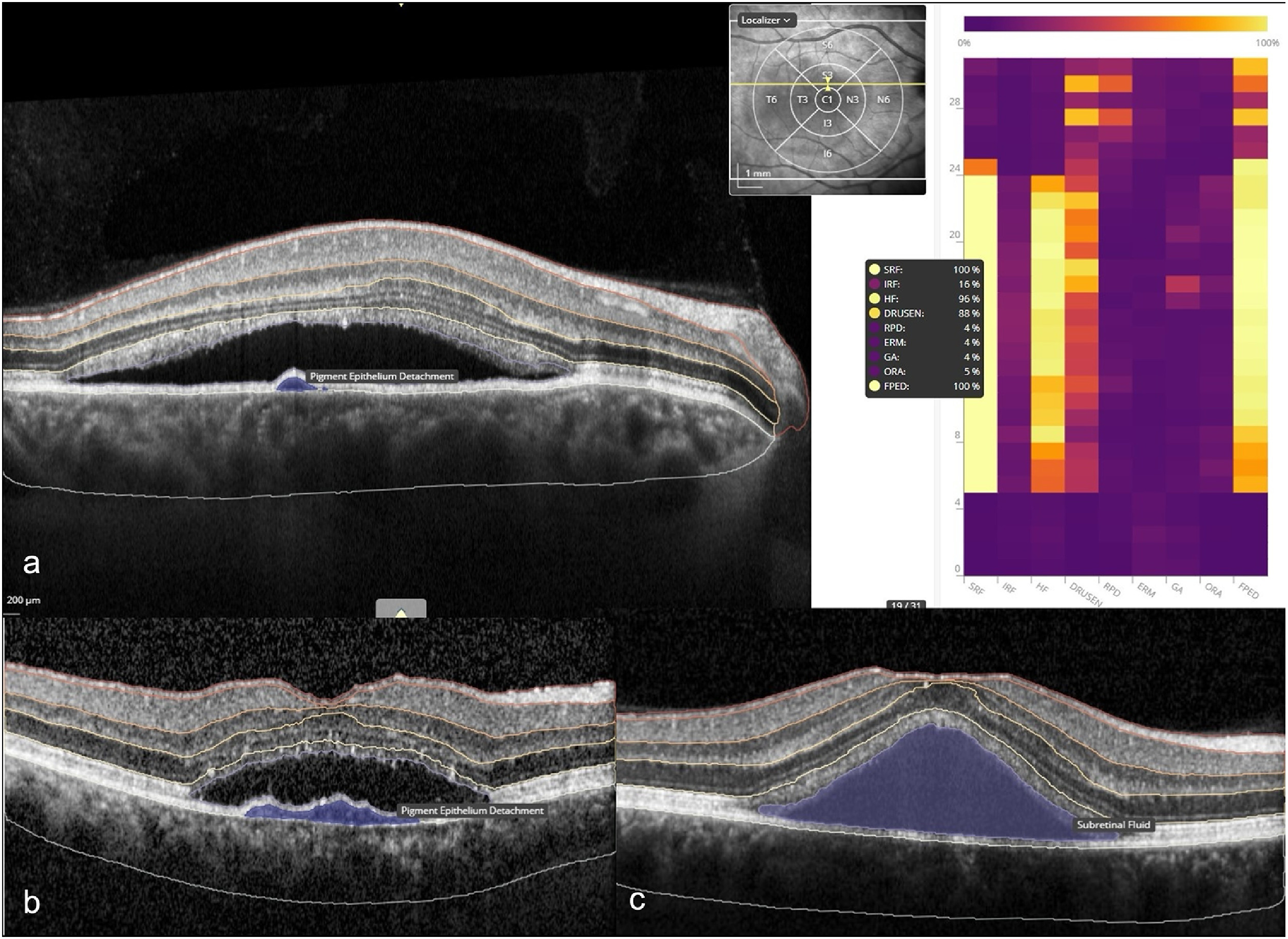
Baseline OCT imaging was analyzed using an AI-based platform (Discovery OCT Biomarker Detector; RetinAI AG), which automatically quantified multiple retinal biomarkers. These included retinal layer thicknesses (notably the outer nuclear layer [ONL] and photoreceptor-retinal pigment epithelium [PR+RPE] complex), SRF and pigment epithelial detachment (PED) fluid volumes, and probabilities of specific OCT features such as flat PED and hyperreflective foci (HF), evaluated across central, parafoveal, and perifoveal ETDRS grid regions.
A Random Forest classifier was developed using these biomarker inputs to predict CSC chronicity at baseline. Model interpretability was enhanced by applying SHapley Additive exPlanations (SHAP) to determine feature importance driving predictions.
Key Findings
Among the cohort, 28% of eyes were classified as acute CSC (with SRF resolution) and 72% as chronic CSC (persistent SRF). Patients with chronic CSC were significantly older (mean 51.7 vs. 43.2 years, p<0.001) and had worse baseline visual acuity (mean logMAR 0.25 vs. 0.16, p=0.029), emphasizing the association between clinical severity and chronicity.
The most strongly associated baseline OCT biomarker with chronic CSC was reduced parafoveal ONL thickness (correlation r = -0.414, q<0.0001), suggesting early outer retinal thinning as an adverse prognostic sign. The Random Forest classifier achieved a robust area under the receiver operating characteristic curve (AUC) of 0.852 (95% CI: 0.748–0.940), demonstrating good discriminative ability in predicting chronic disease from baseline imaging alone.
SHAP analysis elucidated the top predictive features: greater parafoveal ONL thickness, increased perifoveal PR+RPE thickness, and larger central SRF volume were strongly predictive of acute CSC. Conversely, increased retinal nerve fiber layer (RNFL) thickness, larger PED volume, and higher probabilities of flat PED and hyperreflective foci were indicative of chronic CSC.
These findings underscore the prognostic value of quantitative markers of outer retinal integrity, fluid distribution, and RPE dysfunction gleaned from automated OCT analysis. Importantly, the interpretable nature of the model aids clinicians in understanding which imaging features contribute most to predicted outcomes.
Expert Commentary
This study represents an important advancement in applying artificial intelligence to retinal disease prognostication. Previous attempts to predict CSC chronicity relied primarily on qualitative OCT assessments or clinical risk factors. By systematically extracting multidimensional OCT biomarkers and leveraging machine learning, the authors provide a more objective and reproducible tool with high predictive accuracy.
The finding that parafoveal ONL thinning predicts chronicity aligns with pathophysiological insights that photoreceptor loss precedes permanent vision loss in CSC. Additionally, the association of greater PED volume and hyperreflective foci with chronic disease may reflect underlying RPE and choroidal pathology contributing to persistent fluid accumulation.
Limitations include the retrospective design and potential variability in imaging protocols across international centers, although the large and ethnically diverse cohort strengthens generalizability. Prospective validation and integration with clinical variables and multimodal imaging could further enhance predictive performance.
Conclusion
This multicenter study establishes that interpretable machine learning algorithms applied to AI-derived baseline OCT biomarkers can predict the clinical course of CSC with good accuracy. Quantitative features reflecting outer retinal layer integrity, fluid morphology, and RPE dysfunction serve as key determinants of disease chronicity.
These insights facilitate early identification of patients at risk for persistent CSC, supporting personalized counseling and management strategies. Future work should aim to prospectively validate these models and explore their integration into clinical workflows to optimize outcomes for CSC patients.
Funding and ClinicalTrials.gov
Information on study funding sources and clinical trial registration was not provided in the original publication summary.
References
1. Desideri LF, Eldridge N, Berger LE, et al. Interpretable Machine Learning for Course Prediction in Central Serous Chorioretinopathy Using Baseline OCT from a Multicenter Cohort. Am J Ophthalmol. 2026 Oct 8. PMID: 42849862.
2. Daruich A, Matet A, Dirani A, et al. Central serous chorioretinopathy: Recent findings and new physiopathology hypothesis. Prog Retin Eye Res. 2015 Mar;48:82-118.
3. Spaide RF, Klancnik JM Jr, Cooney MJ. Retinal pigment epithelial detachments. Retina. 2018;38(11):2092-2115.
4. Boon CJF, Breukink MB, van Grinsven MJJP, et al. Clinical features and long-term prognosis of chronic central serous chorioretinopathy. Ophthalmology. 2017 Feb;124(2):161-169.
This article was created using several editorial tools, including AI, as part of the process. Human editors reviewed this content before publication.