Highlight
This prospective multicenter study identifies a clinically significant brachial-to-radial arterial pressure gradient (NIBR-APG) in critically ill patients treated with vasoactive drugs. Nearly 20% exhibited a gradient exceeding 11 mm Hg, associated with higher vasoactive dosing, enhanced organ dysfunction, and increased intensive care unit (ICU) mortality.
The study underscores the importance of recognizing noninvasive brachial-to-radial arterial pressure discrepancies during hemodynamic monitoring to optimize vasoactive therapy in critically ill populations.
Study Background
Accurate arterial pressure monitoring is essential in managing critically ill patients receiving vasoactive agents, used to stabilize hemodynamics in shock states. Invasive arterial lines, commonly placed in the radial artery, provide continuous blood pressure measurement. However, peripheral arterial pressure readings may not always reflect central arterial pressure due to physiological and pathological variations, especially under vasopressor influence.
Previous case reports and small series have described a brachial-to-radial arterial pressure gradient, which can lead to underestimation of true blood pressure if relying solely on radial artery measurements. Understanding the prevalence and clinical significance of this gradient in a large critically ill cohort has implications for patient safety and hemodynamic management.
Study Design
This was a prospective observational study conducted across 13 intensive care units in Poland, including adult ICU patients with radial artery catheters receiving vasoactive agents from April to July 2025. A total of 303 patients were enrolled, encompassing both surgical and medical admissions, including a significant subgroup with septic shock.
Invasive radial mean arterial pressure (MAP) was recorded, alongside noninvasive brachial MAP measurements, each taken twice and averaged. A predefined significant noninvasive brachial-to-radial arterial pressure gradient (NIBR-APG) was set at >11 mm Hg. Clinical variables, vasoactive drug dosages converted to norepinephrine equivalents, Sequential Organ Failure Assessment (SOFA) scores, peripheral perfusion indicators, lactate levels, and ICU outcomes were collected and analyzed.
Key Findings
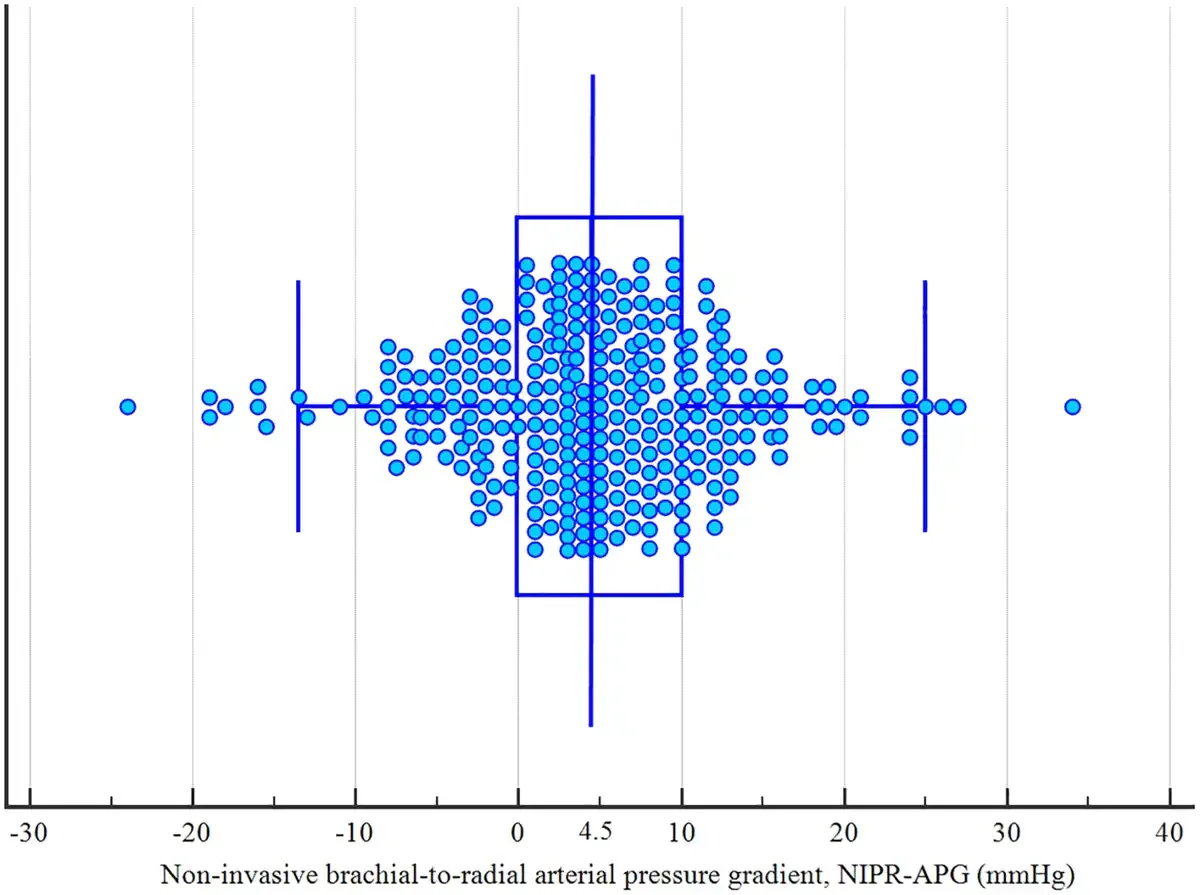
The median NIBR-APG in the cohort was 4.5 mm Hg (interquartile range 0.0–10.0 mm Hg). Notably, 56 patients (18.4%) demonstrated a clinically significant gradient >11 mm Hg, indicating a substantial discrepancy between brachial noninvasive and radial invasive measurements.
Patients with a significant pressure gradient received approximately double the vasoactive drug dose (median 0.23 vs. 0.10 µg/kg/min norepinephrine equivalents, p < 0.01) compared to those without such gradients. This subset also exhibited higher illness severity as reflected by elevated SOFA scores (median 11 vs. 10), prolonged capillary refill times (3.0 vs. 2.5 seconds), and elevated serum lactate concentrations (2.7 vs. 1.3 mmol/L), markers consistent with impaired tissue perfusion and oxygenation.
Multivariate logistic regression identified vasoactive agent dose as the sole independent predictor for NIBR-APG exceeding 11 mm Hg (odds ratio 1.68, 95% CI 1.12–2.52), highlighting a dose-response relationship between vasopressor support intensity and arterial pressure gradient presence.
Of clinical concern, ICU mortality was significantly increased among patients with a pronounced pressure gradient (48.0% vs. 30.1%, p < 0.05), suggesting that these gradients might indicate or contribute to worse outcomes.
Expert Commentary
This study addresses an important yet under-recognized phenomenon in critical care hemodynamic monitoring. The findings suggest that reliance solely on radial artery pressure measurements without corroborating more central or alternative site pressures may lead to underestimation of arterial pressure and inappropriate vasoactive management.
The dose-dependence of the pressure gradient points toward vasoconstriction effects induced by vasoactive agents differentially impacting distal arterial segments. This can cause pressure amplification or dampening distally, complicating clinical interpretation.
Limitations include the observational design without longitudinal follow-up to determine causality or direct impact on outcomes. Also, noninvasive brachial pressure measurements may vary with cuff size and patient factors, potentially affecting gradient calculation.
Current guidelines do not explicitly address arterial site discrepancies; hence, these data advocate for greater vigilance and potentially multi-site pressure monitoring in patients on high-dose vasoactive therapy. Further research is warranted to explore mechanistic pathways and the impact of pressure gradients on titration of vasoactive agents and patient-centered outcomes.
Conclusion
The brachial-to-radial arterial pressure gradient is a common and clinically relevant phenomenon in critically ill patients treated with vasoactive agents. Its presence correlates with increased vasoactive dosing, higher severity of organ dysfunction, and greater ICU mortality.
Recognition of this gradient should prompt critical care clinicians to consider arterial pressure measurement site selection carefully and integrate multiple hemodynamic parameters to guide vasoactive therapy prudently. Improved awareness may lead to more accurate blood pressure assessment and potentially better patient management and outcomes.
Funding and ClinicalTrials.gov
The study was conducted by the Polish Perioperative and Critical Care Research Group (PERI-CRIT) without reported industry funding. The clinical trial registration number was not provided.
References
1. Putowski Z, Gawda R, Czarnik T, et al. Brachial-to-Radial Pressure Gradient in Critically Ill Patients Treated With Vasoactive Agents: A Prospective Observational Study. Crit Care Med. 2026 Oct 23. PMID: 42919451.
2. Osman D, Ridel C, Ray P, et al. Accuracy of invasive arterial pressure monitoring: insights from the analysis of a large database of arterial pressure waveforms. Crit Care Med. 2015;43(12):e542–e544.
3. Rivers E, Nguyen B, Havstad S, et al. Early goal-directed therapy in the treatment of severe sepsis and septic shock. N Engl J Med. 2001 Nov 8;345(19):1368–77.