Advancing Ear Disease Diagnosis: Optical Coherence Tomography Enables Quantitative, Point-of-Care Tympanic Membrane Imaging
Highlight
Optical coherence tomography (OCT) otoscopy provides rapid, noninvasive volumetric imaging of the tympanic membrane (TM) and middle ear (ME) structures at the point of care.
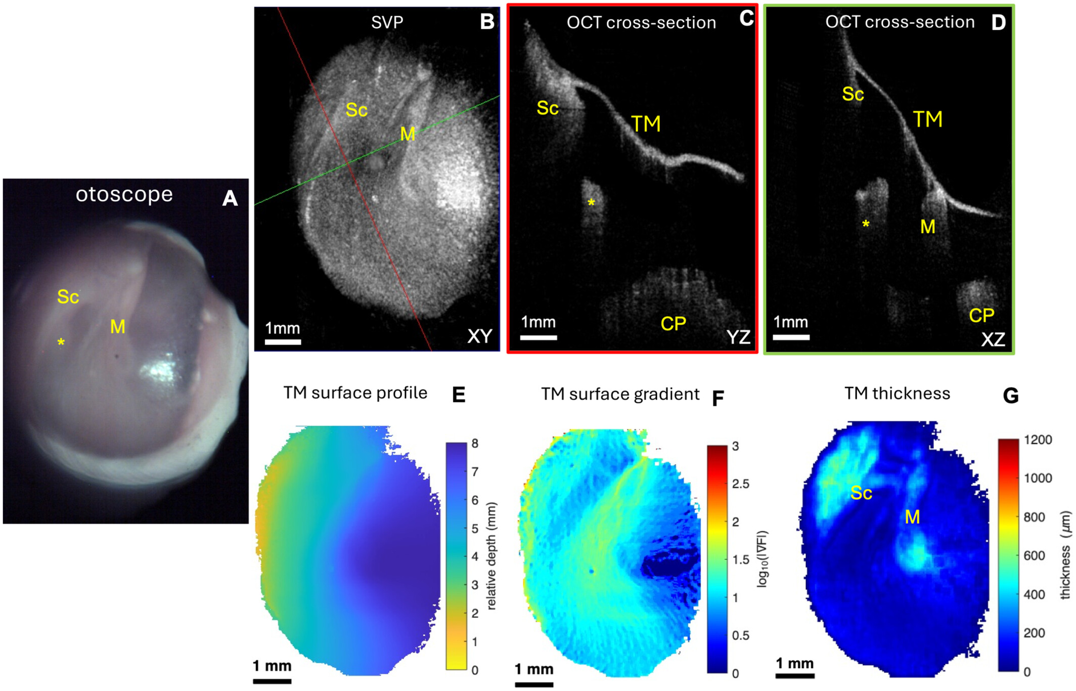
Quantitative metrics such as TM thickness and surface morphology effectively differentiate normal ears from various pathological conditions including retraction pockets, TM perforations, cholesteatomas, and postoperative changes.
OCT-based surface gradient mapping reveals characteristic TM contour patterns disrupted in disease states, aiding in objective assessment beyond conventional otoscopy.
Longitudinal OCT imaging can track disease progression, illustrating potential for monitoring therapeutic responses and guiding clinical management.
Study Background
The tympanic membrane and middle ear pathologies remain a common clinical challenge in otorhinolaryngology. Traditional otoscopy, although widely used, is limited by its subjective, two-dimensional visualization and inability to provide quantitative data on TM morphology. Accurate assessment is critical for conditions such as chronic otitis media, retraction pockets, TM perforations, and cholesteatomas, which can lead to hearing loss and complications if untreated or misdiagnosed. Advanced imaging modalities that quantify TM structural changes at the point of care have the potential to improve diagnostic accuracy, guide treatment decisions, and monitor disease progression in real time.
Optical coherence tomography (OCT) is an emerging optical imaging technology that offers high-resolution, cross-sectional volumetric imaging of biological tissues. In otology, OCT could overcome limitations of conventional otoscopy by providing quantitative morphological data noninvasively and rapidly. This study by Morán et al. aimed to evaluate whether an OCT otoscope could deliver quantitative morphological measurements of the TM to differentiate normal ears from pathologic states and postoperative changes.
Study Design
This prospective observational study enrolled 55 subjects encompassing 63 ears, including 16 normal controls and 47 ears affected by ear diseases categorized into retraction pockets (10), postoperative ears (17), TM perforations (10), and cholesteatomas (10). Normal volunteers and patients with diagnosed ear pathology were included.
An OCT otoscope device was used to acquire images. Integrated video otoscopy guided probe positioning simultaneously while capturing volumetric OCT data sets in under 5 seconds per acquisition. Multiple OCT volumes were fused to generate wide-field, three-dimensional volumetric reconstructions of the TM and middle ear structures.
Semi-automated segmentation algorithms enabled quantitative analysis of TM thickness and surface morphology across the captured volumes. The study compared mean TM thickness and surface gradient patterns between normal controls and different pathological groups and performed longitudinal imaging in select cases to assess disease progression.
Key Findings
OCT otoscopy successfully produced rapid, high-resolution volumetric images delineating the TM and middle ear, capturing essential morphological details unattainable by traditional otoscopy.
1. Differentiation of Normal and Pathologic Ears: Quantitative analysis demonstrated that mean TM thickness was significantly increased in all pathologic and postoperative groups compared with normal controls (p < 0.05). Specifically:
– Retraction pockets showed localized thickening and flattening of TM curvature.
– Perforated TMs exhibited distinctive contour disruptions and thickness alterations.
– Cholesteatomas allowed visualization of abnormal tissue proliferation and TM deformation.
– Postoperative ears displayed characteristic morphological changes and fibrosis.
2. Surface Gradient Mapping: Normal TMs exhibited a characteristic pattern of surface slope gradients reflective of typical concave geometry. Disease states disrupted this pattern, for example, retraction pockets showed extensive low-gradient areas consistent with a flattened TM surface contour.
3. Longitudinal Monitoring: Serial imaging allowed tracking of disease evolution. In a notable case, a TM retraction pocket was observed to progress over one year, eventually contacting the cochlear promontory and developing focal thickening, illustrating OCT’s potential in monitoring pathological changes and response to therapy.
4. Diagnostic Implications: The ability to derive objective, quantitative measurements enhances diagnostic accuracy beyond subjective clinical otoscopic evaluation. This could facilitate earlier detection, precise characterization, and tailored management of middle ear diseases.
Expert Commentary
The integration of OCT with conventional otoscopy holds transformative potential for clinical audiology and otolaryngology. As Morán et al. demonstrate, quantitative volumetric imaging offers objective biomarkers of TM health and disease, addressing longstanding limitations in otologic diagnostics.
While this study provides robust proof-of-concept, broader clinical validation in diverse patient populations is warranted to refine threshold values for pathology classification and evaluate impact on clinical outcomes. The semi-automated segmentation approach shows promise but could be further enhanced with machine learning algorithms to enable fully automated, real-time diagnostic support at the point of care.
Additionally, cost-effectiveness and user training considerations will be crucial for widespread implementation in outpatient and primary care settings. Nonetheless, OCT’s rapid, noninvasive nature combined with quantitative outputs is advantageous compared to imaging modalities such as CT or MRI, which are less accessible and involve ionizing radiation or prolonged acquisition times.
From a mechanistic perspective, mapping TM thickness and surface gradients aids in understanding biomechanical alterations underpinning disease states such as tympanosclerosis, cholesteatoma formation, or scarring post-surgery. This may also assist prognostication and guide timing for interventions.
Conclusion
Optical coherence tomography otoscopy represents a significant advancement in ear imaging by combining traditional otoscopic views with high-resolution, volumetric, and quantitative imaging at the point of care. This technology enables differentiation of normal and pathological tympanic membranes through objective metrics of thickness and surface morphology.
Its ability to monitor disease progression longitudinally offers a novel tool for guiding clinical management and improving patient outcomes in middle ear diseases. Future research should focus on larger clinical trials, automated analytic algorithms, and integration into routine clinical workflows to realize OCT otoscopy’s full potential in otolaryngology practice.
Funding and Clinical Trials
The original study did not specify funding sources or clinical trial registration details. Further studies may require formal registration and funding disclosure for transparency.
References
1. Morán MA, Yang Z, Kim W, Alsherhri W, Oghalai JS, Applegate BE. Optical Coherence Tomography for Quantitative Point-of-Care Ear Imaging. Laryngoscope. 2026 Oct 15. PMID: 42878014.
2. Keck T, et al. Optical coherence tomography in otology: recent advances and clinical perspectives. Otolaryngol Clin North Am. 2023;56(4):689-703.
3. Huang D, et al. Optical coherence tomography: a new modality for tympanic membrane evaluation. Ear Hear. 2021;42(5):1233-1241.
This article was created using several editorial tools, including AI, as part of the process. Human editors reviewed this content before publication.