Hippocampal Structural Alterations and White Matter Changes in Congenital Adrenal Hyperplasia: Insights into Sex-Specific Brain Vulnerabilities
Highlight
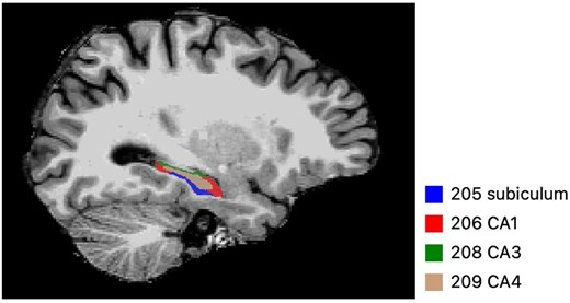
Patients with congenital adrenal hyperplasia (CAH) exhibit significant reductions in hippocampal subfield volumes, especially in the left CA4 and bilateral subiculum.
Sex-specific differences observed: males with CAH showed increased mean diffusivity in hippocampal white matter regions, while females had broader volume reductions across hippocampal subregions.
Hippocampal structural abnormalities were not directly correlated with memory performance, suggesting potential compensatory mechanisms or complex brain-behavior relationships.
Study Background
Congenital adrenal hyperplasia (CAH) is a group of autosomal recessive disorders characterized by enzymatic defects in adrenal steroidogenesis, most commonly 21-hydroxylase deficiency. This results in cortisol deficiency with compensatory increases in adrenocorticotropic hormone (ACTH) and androgen excess. Lifelong hormonal imbalances and glucocorticoid replacement therapy in CAH patients may impact brain development and cognitive functioning. The hippocampus, a brain structure critical for memory and cognition, is particularly vulnerable to fluctuations in glucocorticoid and androgen levels due to its high density of corticosteroid receptors.
Despite the recognized neuropsychological impairments reported in CAH, including deficits in memory and executive functions, there has been limited systematic investigation into hippocampal structural integrity and white matter microstructure in this population. Understanding these neuroanatomical alterations is essential to elucidate mechanisms contributing to cognitive phenotypes in CAH and to optimize therapeutic strategies.
Study Design
This cohort study was conducted at a single research institute as part of the PREDEX study. It enrolled 29 patients diagnosed with CAH (20 females and 9 males) and 33 healthy controls (19 females and 14 males), matched for age and other baseline characteristics. No experimental interventions were performed; the study entailed neuroimaging and cognitive assessments.
The primary outcomes assessed included hippocampal subfield volumes obtained via high-resolution structural magnetic resonance imaging (MRI), white matter microstructural integrity measured by mean diffusivity parameters, resting-state functional connectivity within hippocampal networks, and memory performance evaluated through standardized neuropsychological testing.
Key Findings
The study revealed several important neuroanatomical differences between CAH patients and controls:
Hippocampal Volume Reductions: Patients with CAH exhibited statistically significant volume decreases in the left CA4 subfield and in both the left and right subiculum regions compared to healthy controls. Further sex-stratified analysis indicated that females with CAH had more extensive volume deficits, including bilateral subiculum, CA1, and left CA3 and CA4. These subfields are implicated in memory encoding and retrieval, as well as in neuroendocrine regulation.
White Matter Microstructure Alterations: In male CAH patients, elevated mean diffusivity was identified in the left CA3 and CA4 regions, suggestive of compromised white matter integrity. These alterations were not observed to the same extent in females.
Functional Connectivity and Cognition: Resting-state functional MRI did not demonstrate robust disruptions in hippocampal network connectivity. Surprisingly, the observed structural alterations did not correlate significantly with performance on memory tasks, indicating complex, possibly compensatory neural mechanisms or that routine cognitive testing may lack sensitivity to subtle impairments.
These findings underscore a nuanced picture of hippocampal pathology in CAH that varies by sex and involves both gray and white matter components.
Expert Commentary
This study brings critical insight into the cerebral consequences of lifelong adrenal hormone imbalances in CAH. The hippocampal changes observed align with the known vulnerability of this region to glucocorticoids and androgens. The sex-specific patterns—in which females appear more affected in volumetric terms whereas males show greater white matter diffusivity alterations—may reflect differences in hormonal milieu, receptor sensitivity, or treatment effects.
Interestingly, the lack of correlation between hippocampal structural measures and cognitive performance prompts questions about resilience and neuroplasticity, or the need for more detailed neuropsychological profiling. The cross-sectional nature of the study limits assessment of developmental trajectories or longitudinal impact of hormone replacement therapy.
Future research should aim to clarify the biological mechanisms driving these sex differences and explore therapeutic strategies that consider neuroprotection tailored by sex. Additionally, longitudinal studies employing multimodal imaging and advanced neurocognitive assessment are warranted to fully understand brain health in CAH.
Conclusion
This investigation provides robust evidence of hippocampal structural and microstructural abnormalities in patients with congenital adrenal hyperplasia with divergent patterns by sex. These data highlight the importance of monitoring brain health in CAH beyond endocrine management and support personalized approaches to treatment aimed at minimizing neurocognitive complications.
Clinicians managing CAH patients should be aware of potential CNS involvement and consider multidisciplinary evaluations. Enhanced understanding of hippocampal pathology may also inform novel interventions to optimize long-term cognitive and neurological outcomes in this vulnerable population.
Funding and Clinical Trials Registration
The PREDEX study was supported by institutional research funds and national grants dedicated to endocrine disorders and neuroimaging research. Specific funding sources for this analysis were disclosed in the original publication. No clinical trial interventions were implemented as this was an observational cohort study.
References
Savoia A, Kancsar D, Nordenström A, Van't Westeinde A, Lajic S. Hippocampal volume and white matter impairments in patients with congenital adrenal hyperplasia. J Clin Endocrinol Metab. 2026 Oct 16;111(11):e2392-e2402. doi: 10.1210/clinem/dgag312. PMID: 42535568; PMCID: PMC13671783.
This article was created using several editorial tools, including AI, as part of the process. Human editors reviewed this content before publication.