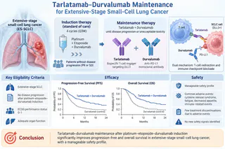
Abstract Intraocular metastases from small-cell lung cancer (SCLC) predominantly affect the choroid, whereas isolated retinal involvement is exceedingly rare and can closely mimic infectious retinitis. We report the case of a 52-year-old man with extensive-stage SCLC who presented with progressive vision loss in the left eye. Initial clinical findings and low-level cytomegalovirus (CMV) viremia prompted empiric antiviral therapy for presumed CMV retinitis. However, lack of clinical response, negative aqueous humor viral nucleic acid amplification testing (NAAT), and marked retinal thickening on optical coherence tomography (OCT) and ocular ultrasonography led to a diagnostic vitrectomy and retinal biopsy, which confirmed metastatic SCLC.
Case Presentation
A 52-year-old man presented to the ophthalmology clinic at Massachusetts General Hospital with a 3-week history of worsening blurred vision, floaters, intermittent ocular pain, and new-onset temporal peripheral vision loss in his left eye.
Twenty-seven months prior to presentation, the patient had been diagnosed with extensive-stage small-cell lung cancer (SCLC) with hepatic metastases. He was initially treated with carboplatin and etoposide, with atezolizumab added 1 month later, followed by maintenance atezolizumab. Over the subsequent two years, his disease course was complicated by metastases to the left adrenal gland (treated with stereotactic body radiotherapy), multiple hemorrhagic brain metastases (treated with whole-brain radiotherapy), and cervical lymphadenopathy, necessitating second-line lurbinectedin and later third-line irinotecan. Six months before his current ophthalmic visit, he experienced seizures, aphasia, and right-sided weakness due to supratentorial progression, which was managed with stereotactic radiotherapy, levetiracetam, and oral dexamethasone. His medical history was also notable for atrial fibrillation, diabetes mellitus, thoracic compression fracture, depression, and a 38-pack-year smoking history.
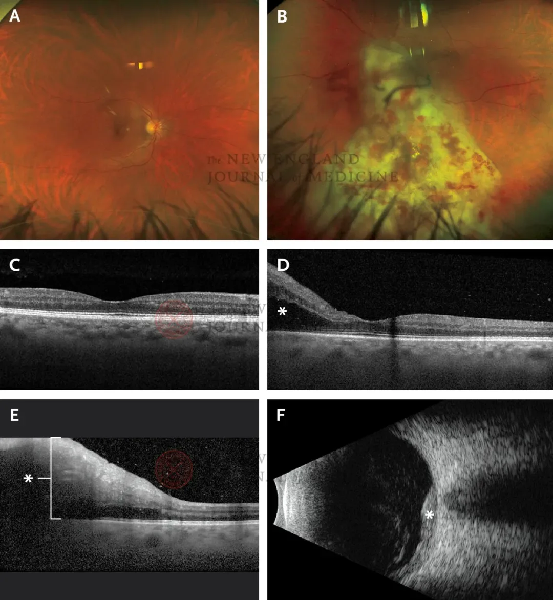
On ophthalmologic examination, visual acuity was 20/100 in the right eye and 20/80 in the left eye. Intraocular pressures and pupillary light reflexes were normal, though confrontational visual field testing revealed an inferotemporal defect in the left eye. Slit-lamp examination showed bilateral cataracts without anterior segment inflammation. Dilated fundus examination of the left eye demonstrated 2+ vitreous cells and a large, wedge-shaped area of retinal whitening accompanied by intraretinal hemorrhages and scant hard exudates extending from the inferior optic disc margin toward the inferotemporal periphery. The right fundus was unremarkable.
Optical coherence tomography (OCT) of the left eye revealed intraretinal fluid nasal to the macula and diffuse intraretinal thickening at the junction between normal and affected retina. Ocular ultrasonography confirmed pronounced retinal thickening in the area of the lesion. Laboratory evaluation showed mild anemia (hemoglobin, 11.9 g/dL), normal leukocyte and platelet counts, and negative HIV-1/2 serologies.
Based on the appearance of the retinal lesion and his history of immunosuppressive cancer therapies, atypical cytomegalovirus (CMV) retinitis was suspected. Anterior chamber paracentesis was performed for viral nucleic acid amplification testing (NAAT), and empiric treatment was initiated with intravitreal foscarnet and oral valganciclovir. Two days later, laboratory results revealed a CD4+ T-cell count of 239/μL and low-level blood CMV DNA viremia (348 IU/mL). Despite biweekly intravitreal foscarnet injections over 2 weeks, the retinal lesion showed no regression. Furthermore, blood CMV DNA declined to 47 IU/mL, CMV IgM was negative (IgG positive), and aqueous humor NAAT returned negative for CMV, herpes simplex virus (HSV), and varicella-zoster virus (VZV) DNA.
Diagnostic Workup and Pathological Findings
Given the absence of clinical response to antiviral therapy, negative aqueous viral testing, and atypical ultrasound findings, an intraocular malignancy was suspected. By this time, visual acuity in the left eye had deteriorated to counting fingers. The patient underwent a diagnostic pars plana vitrectomy combined with a retinal biopsy.
Histopathological examination of the paraffin-embedded retinal biopsy specimen demonstrated sheets of high-grade, mitotically active, primitive small round blue cells with scant cytoplasm, pleomorphic nuclei, finely granular ("salt-and-pepper") chromatin, and focal nuclear molding, without true rosettes. Immunohistochemical staining was strongly positive for pancytokeratin (AE1/AE3/CAM5.2, showing characteristic dot-like and perinuclear ring patterns) and synaptophysin. Cytologic analysis of the concurrent vitreous specimen similarly revealed atypical hyperchromatic round cells amid inflammatory and apoptotic debris. These findings confirmed a diagnosis of high-grade neuroendocrine carcinoma consistent with retinal metastasis from the patient’s primary SCLC.
Treatment and Outcome
Following histological confirmation, the patient was referred to ocular oncology. Examination of the left eye showed progression to no light perception, an elevated intraocular pressure of 28 mm Hg, and clinically significant anterior chamber cells, indicating intraocular tumor dissemination and probable optic nerve infiltration. Orbital magnetic resonance imaging (MRI) demonstrated posterolateral nodular enhancement of the left globe and mild enhancement of the intraocular segment of the left optic nerve.
After discussing management options—including observation, systemic chemotherapy, local radiation, and enucleation—the patient elected palliative globe-sparing radiotherapy. He received volumetric modulated arc therapy (VMAT) to the entire left globe at a total dose of 20 Gy delivered in 5 fractions. Prior to his scheduled 6-week follow-up visit, the patient returned to Egypt, where he subsequently died from complications related to systemic progression of SCLC.
Discussion
Ocular metastasis occurs in approximately 2% to 9% of patients with systemic malignancies, with lung and breast cancers representing the most common primary sources. Within the eye, the uveal tract—specifically the highly vascular choroid—is overwhelmingly favored, whereas isolated retinal metastasis accounts for less than 1% of all intraocular metastases. Because SCLC has a strong propensity for central nervous system dissemination, concurrent brain metastases (as seen in this patient) serve as a critical clinical clue for intraocular spread.
This case highlights a significant diagnostic pitfall in immunocompromised oncology patients. Low-level CMV DNAemia is relatively common following chemotherapy and corticosteroid administration, yet it rarely correlates with true end-organ CMV retinitis in HIV-negative individuals. Reliance on peripheral blood viral load alone can delay accurate diagnosis. Key features arguing against CMV retinitis in this patient included the negative aqueous humor CMV NAAT (which carries a sensitivity exceeding 90% in true CMV retinitis), a moderately preserved CD4+ T-cell count (239/μL), lack of response to targeted antiviral therapy, and marked retinal thickening on OCT and B-scan ultrasonography—a structural hallmark more characteristic of neoplastic infiltration than viral necrosis.
When clinical and imaging features diverge from infectious etiologies, prompt vitrectomy with retinal biopsy is warranted to establish a definitive tissue diagnosis and guide timely palliative radiotherapy, which can preserve the globe and alleviate ocular symptoms.
Reference
Sobrin L,McKay KM,and Kovac V,et al. Case 30-2026:A 52-Year-Old Man with Vision Loss in the Left Eye. N Engl J Med 2026;395:1635-1641