Introduction
Progressive pulmonary fibrosis (PPF) is characterized by progressive scarring of lung tissue that leads to a decline in lung function and ultimately respiratory failure. Monitoring disease progression and treatment response accurately is essential for improving patient outcomes. Forced vital capacity (FVC) is the commonly used clinical endpoint in trials, yet it has known limitations in sensitivity and an inability to capture nuanced structural changes. To address this, quantitative computed tomography (QCT) imaging is emerging as a powerful tool to detect and predict progression in PPF more sensitively and objectively.
The INBUILD trial, a double-blind, randomized, placebo-controlled study, evaluated the efficacy of nintedanib, an antifibrotic agent, in patients with PPF across various underlying interstitial lung diseases (ILD). A sub-study within INBUILD focused on using QCT methods to refine prognostication and assess treatment effects.
Quantitative CT Methods: UCLA and e-Lung Algorithms
Two specialized algorithms were used to derive QCT measures from lung scans:
The University of California Los Angeles (UCLA) algorithm generates quantitative ILD (QILD) and quantitative lung fibrosis (QLF) scores. QILD encompasses the total extent of interstitial abnormalities, whereas QLF specifically quantifies fibrotic changes.
The e-Lung (Brainomix) algorithm calculates total disease extent (TDE), reticulovascular score (RVS), and weighted RVS representing different components of lung pathology.
These metrics aim to objectively quantify the amount and severity of fibrotic and other pathological changes in lung tissue, allowing numerical tracking of disease burden over time.
Study Design and Patient Population
The sub-study included 474 patients with PPF enrolled in the INBUILD trial. Patients had varying ILD diagnoses but demonstrated progressive fibrosis as defined by clinical and radiological criteria. Participants were randomized to receive either placebo or nintedanib and underwent serial lung function testing and CT imaging over 52 weeks.
Key Findings
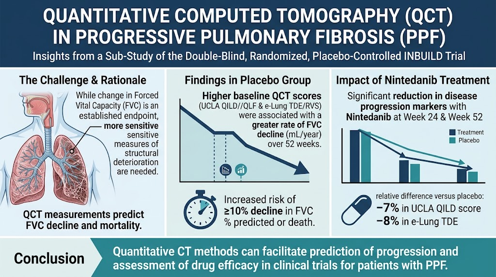
Baseline QCT Scores and Lung Function Decline: Among placebo patients, higher baseline QILD, QLF, and e-Lung scores correlated with a more rapid decline in FVC over one year. This relationship supports QCT measures as predictive markers of disease progression beyond what baseline FVC alone can provide.
Prognostic Value for Clinical Outcomes: Patients with QCT scores above median values had an increased risk of a ≥10% decline in predicted FVC or death over 52 weeks compared to those below the median. This risk translated to a shorter restricted mean survival time by approximately 40 to 65 days, emphasizing the clinical relevance of these imaging biomarkers.
Effects of Nintedanib Treatment: Nintedanib significantly reduced progression as measured by changes in UCLA QILD scoring and e-Lung TDE at both 24 and 52 weeks compared with placebo. The relative differences ranged between 7% and 8%, indicating treatment slowed fibrotic changes detectable by sensitive QCT methods.
Clinical Implications
These results demonstrate that quantitative CT imaging can be a valuable biomarker in clinical practice and research. By providing a more detailed and reproducible assessment of lung fibrosis extent and progression, QCT complements traditional lung function tests.
In clinical trials, QCT metrics can enhance endpoint sensitivity to detect treatment effects, reducing required sample sizes and study durations. For clinicians, QCT offers a potential means to better stratify patients according to risk and tailor monitoring and therapeutic strategies accordingly.
Limitations and Future Directions
While promising, QCT techniques require continued validation across diverse populations and ILD subtypes. Standardization of imaging acquisition and processing protocols remains essential for broader adoption.
Further research integrating QCT with clinical data and novel biomarkers may improve individualized prognosis and therapeutic decision-making. Additionally, long-term studies are needed to understand the relationship between QCT changes and survival or quality of life outcomes.
Conclusion
Quantitative CT imaging, using the UCLA and e-Lung algorithms, provides robust prognostic information and sensitivity to treatment response in progressive pulmonary fibrosis. These methods represent important tools for advancing clinical research and enhancing patient management in this challenging disease.
Expanded Context on Nintedanib and PPF Management
Nintedanib is a tyrosine kinase inhibitor that has demonstrated efficacy in slowing fibrosis progression in idiopathic pulmonary fibrosis (IPF) and PPF from diverse causes by targeting multiple pathways involved in fibrotic tissue remodeling. It is administered orally and generally well-tolerated, with common side effects including gastrointestinal symptoms. Its approval for PPF reflects a shift towards treating the fibrosis phenotype regardless of the underlying ILD etiology.
Management of progressive pulmonary fibrosis remains complex, often involving multidisciplinary teams including pulmonologists, radiologists, and rheumatologists. Timely diagnosis and tracking of progression with tools like QCT alongside physiological measures enable better prognostication and early therapeutic intervention, which is critical to preserving lung function and improving patient quality of life.
Reference
Devaraj A, George PM, Joly O, Goldin J, White ES, Ittrich C, Stowasser S, Risse F. Quantitative Computed Tomography in Progressive Pulmonary Fibrosis: Data from a Sub-Study of the Double Blind, Randomized, Placebo-controlled INBUILD Trial. Am J Respir Crit Care Med. 2026 Sep 29:aamag526. doi: 10.1093/ajrccm/aamag526. Epub ahead of print. PMID: 42809243.