Highlight
EB-OCT enables large-volume, microscopic-resolution imaging of lung tissue, providing detailed ILD-specific features non-invasively.
In a cohort of 15 patients with difficult-to-diagnose ILD, EB-OCT showed 100% agreement with final multidisciplinary diagnosis, including cases where TBLC was non-diagnostic.
Combining EB-OCT with TBLC and Envisia Genomic Classifier augmented diagnostic confidence and refined differential diagnoses consistently across independent multidisciplinary centers.
This pilot study supports EB-OCT as a promising complementary modality to existing ILD diagnostic tools, warranting multicenter validation for broader clinical adoption.
Study Background
Interstitial lung diseases (ILDs) constitute a heterogeneous group of pulmonary disorders characterized by varying degrees of inflammation and fibrosis. Accurate diagnosis is critical due to therapeutic and prognostic implications, particularly for idiopathic pulmonary fibrosis (IPF) and other forms of usual interstitial pneumonia (UIP). Traditional diagnostic approaches often rely on surgical lung biopsy (SLB), the gold standard for histopathologic diagnosis, but SLB is invasive and carries significant morbidity risks, limiting its clinical applicability.
Emerging less-invasive diagnostic strategies include transbronchial lung cryobiopsy (TBLC) and molecular classifiers like the Envisia Genomic Classifier (EGC). TBLC offers tissue sampling with fewer complications than SLB but may provide limited sample size and yield. EGC uses genomic profiling to categorize ILD into UIP or non-UIP patterns but reduces diagnostic complexity to binomial outcomes, potentially oversimplifying heterogeneous disease presentations.
Endobronchial optical coherence tomography (EB-OCT) represents an innovative bronchoscopic imaging technique providing high-resolution, cross-sectional images at a microscopic scale, enabling visualization of lung microarchitecture over relatively large volumes. Prior research suggests that EB-OCT can identify morphological features correlating with ILD subtypes, potentially serving as a safer, adjunctive diagnostic modality.
Study Design and Methods
This pilot prospective study enrolled 15 patients clinically referred for TBLC with EGC due to low-confidence ILD diagnoses. These subjects underwent concurrent EB-OCT imaging during bronchoscopy. Each diagnostic modality—EB-OCT, TBLC histopathology, and EGC genomic classification—was interpreted independently and in a blinded manner by expert pathologists.
Two independent multidisciplinary discussions (MDDs) were convened to finalize ILD diagnoses: one at a center experienced with EB-OCT and another without prior EB-OCT expertise, ensuring reproducibility across settings. The primary endpoint was the concordance of EB-OCT findings with the final consensus diagnosis established by MDD integrating all clinical, radiologic, and conventional biopsy data.
Key Findings
The 15 patients had final diagnoses including UIP/IPF (n=5), secondary UIP (n=2), and diverse non-UIP ILDs (n=8), with full agreement between the two MDD groups.
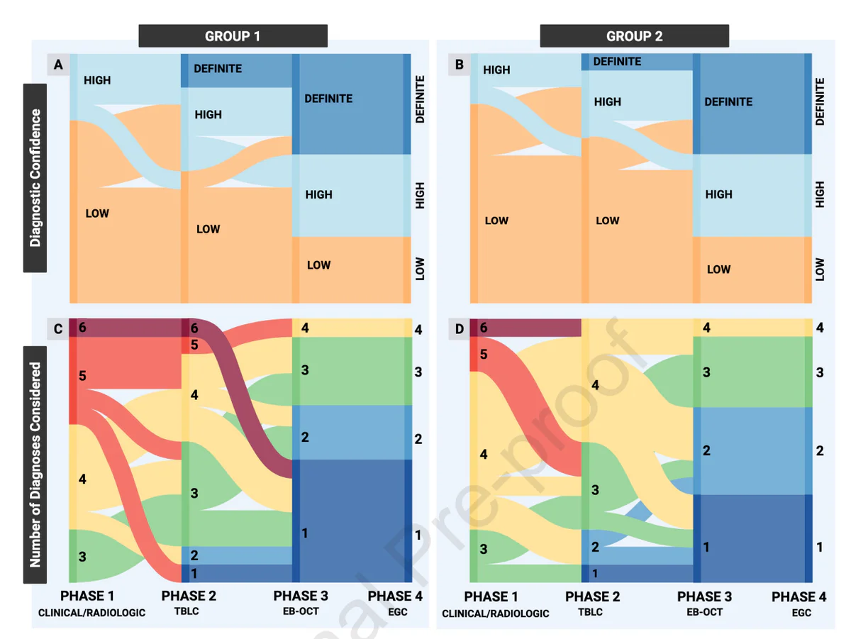
Most notably, EB-OCT demonstrated diagnostic data concordant with the final MDD diagnosis in 100% of cases (15/15). This encompassed six patients (40%) where TBLC was non-diagnostic, highlighting EB-OCT’s potential to provide critical diagnostic information when tissue sampling fails. For the nine patients with diagnostic TBLC results (60%), EB-OCT findings were fully concordant.
Combining EB-OCT with TBLC/EGC enhanced diagnostic confidence and reduced diagnostic ambiguity in all patients compared to initial clinical and radiologic assessments. Importantly, these improvements were independently replicated in both MDD centers, supporting generalizability.
Safety was not explicitly detailed in the abstract, but the low-risk bronchoscopic nature of EB-OCT suggests a favorable procedural profile compared to more invasive biopsies.
Expert Commentary
This study addresses a critical unmet need in ILD diagnostics: balancing the demand for comprehensive histopathologic evaluation with patient safety. EB-OCT’s ability to perform large-volume, microscopic-resolution imaging complements TBLC’s limited sampling and EGC’s genomic binary classifier, offering clinicians a richer, multimodal dataset during diagnosis.
The 100% concordance rate with MDD diagnosis, including cases where TBLC was non-diagnostic, suggests EB-OCT may reduce reliance on surgical biopsy and improve diagnostic pathways. Moreover, consistent results across independent MDD panels, including those unfamiliar with EB-OCT, support its reproducibility and ease of integration.
However, this study’s small sample size and single-center origin limit broad inference. Further multicenter studies with larger cohorts are necessary to validate findings, define operational protocols, and evaluate impact on clinical outcomes and healthcare resource use.
Additionally, integration of EB-OCT into clinical workflows demands training and experience to optimize image interpretation and pattern recognition. Future efforts should focus on standardizing nomenclature and developing automated analysis tools to enhance objetivity.
Conclusion
Endobronchial optical coherence tomography represents a promising adjunctive imaging modality in the multidisciplinary diagnosis of interstitial lung disease. This pilot study demonstrates that EB-OCT provides detailed, large-volume microscopic structural information that agrees with final expert consensus diagnosis, enhances diagnostic confidence, and complements existing bronchoscopic biopsy and molecular tools.
Incorporation of EB-OCT into ILD diagnostic algorithms has the potential to reduce invasive procedures, expedite diagnosis, and improve patient-specific management. Nonetheless, validation in larger, multicenter trials remains imperative before routine clinical application.
Funding and ClinicalTrials.gov
Funding sources were not specified in the abstract. Registration details, if existing, were not provided.
References
Arao RK, Yamamoto S, Nandy S, Sharma A, Lee J, Berigei SR, et al. Diagnostic Contribution of Endobronchial Optical Coherence Tomography in the Multidisciplinary Diagnosis of Interstitial Lung Disease. Chest. 2026 Oct 22. PMID: 42913881.
Other relevant references for context (not in original abstract):
– Raghu G, et al. Diagnosis of idiopathic pulmonary fibrosis. An official ATS/ERS/JRS/ALAT clinical practice guideline. Am J Respir Crit Care Med. 2018.
– Ikeda N, et al. Transbronchial lung cryobiopsy in interstitial lung disease: review of procedure, complications, and diagnostic utility. Respir Investig. 2020.
– Maldonado F, et al. The Envisia Genomic Classifier for Usual Interstitial Pneumonia: A Multicenter Validation Study. Am J Respir Crit Care Med. 2019.
These additional references underscore the evolving landscape of less-invasive ILD diagnostic modalities.