Highlight
This prospective study demonstrates that a real-time ultrasound-assisted laryngeal electromyography (LEMG) protocol using a single in-plane lateral oblique trajectory for needle guidance is feasible and safe. The technique achieved 100% electromyography-confirmed muscle sampling success of the thyroarytenoid (TA) and cricothyroid (CT) muscles in elderly patients, despite prevalent thyroid cartilage calcification. Ultrasound allowed effective trajectory guidance and vascular avoidance without the need for electromagnetic tracking equipment.
Study Background
Laryngeal electromyography (LEMG) is a diagnostic tool to evaluate suspected neuromuscular dysfunction of laryngeal muscles, typically employed in patients with voice disorders, vocal fold paralysis, or neuromuscular disease affecting phonation. Precise needle placement into targeted laryngeal muscles such as the thyroarytenoid and cricothyroid is crucial for accurate electromyographic signal acquisition. Conventional LEMG techniques largely rely on landmark-based guidance, which may be technically challenging and can be limited by anatomical variability, especially in elderly patients with cartilage calcifications.
Ultrasound guidance can potentially improve needle targeting accuracy and procedural safety by real-time visualization of anatomical structures. However, existing ultrasound-assisted LEMG protocols often require complex electromagnetic tracking equipment or multiple scanning views, which may not be widely accessible. This study explores a simplified approach using a single dedicated in-plane trajectory ultrasound view to guide LEMG needle insertion in a real-time setting without additional tracking devices.
Study Design
This was a prospective single-arm feasibility study conducted at a single university hospital’s department of rehabilitation medicine. Sixteen consecutive adult patients (14 men and 2 women, mean age 69.5 ± 10.9 years) referred for LEMG due to suspected neuromuscular laryngeal dysfunction were enrolled.
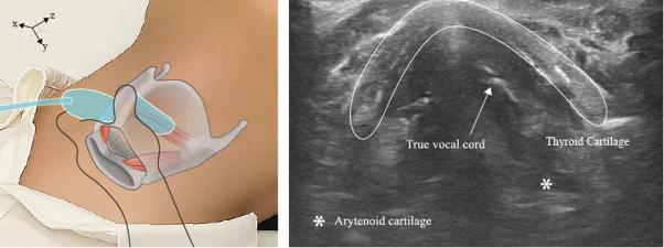
Two physicians performed the procedures using an identical standardized ultrasound-guided protocol. Following an anatomic survey with three orientation views to identify key landmarks, a single in-plane lateral oblique trajectory was used to sample the thyroarytenoid and cricothyroid muscles. Needle insertion was guided by real-time ultrasound without electromagnetic tracking.
Main outcome measures included: (i) electromyography-confirmed muscle sampling success; (ii) completeness of real-time needle visualization during insertion; (iii) ultrasound visualization rates of laryngeal structures (thyroid cartilage, arytenoid, vocal cords, muscles); and (iv) procedure-related adverse events.
Key Findings
A total of 44 muscles were sampled, comprising 31 thyroarytenoid and 13 cricothyroid muscles. The electromyography-confirmed successful sampling rate was 100% (44/44 muscles).
Complete real-time needle visualization was achieved in all cricothyroid muscles (13/13, 100%), while it was limited in the thyroarytenoid muscles, with complete visualization achieved in only 4/31 cases (12.9%). High prevalence of thyroid cartilage calcification (noted in 87.5% of patients) was identified as the principal factor limiting needle visibility in the TA muscle region.
Ultrasound visualized the TA and CT muscles in all cases (100%). The arytenoid and vocal cords were visualized in 93.8% (30/32) of examinations.
Adverse events were minimal: transient coughing occurred in 6.8% (3/44) of muscle examinations but did not necessitate procedure termination. Importantly, no major complications such as bleeding, hematoma, or infection were reported.
Expert Commentary
This feasibility study validates that real-time ultrasound-assisted LEMG using a single in-plane trajectory approach is practical and safe for clinical use. The approach leverages commodity ultrasound equipment without reliance on complex electromagnetic tracking, enhancing accessibility.
The limitation in needle visualization for the TA muscle principally due to thyroid cartilage calcification underscores the challenge in elderly populations. Nevertheless, ultrasound provides critical guidance on the needle trajectory and helps avoid vascular structures, with electromyographic signals remaining the gold standard for final needle localization confirmation.
Further research, ideally a comparative randomized controlled trial against conventional landmark-guided LEMG techniques, is indicated to rigorously assess the added procedural accuracy, patient comfort, cost-effectiveness, and clinical outcomes.
Clinicians should consider the patient’s age-related anatomical changes when employing ultrasound guidance and combine ultrasound with EMG signal interpretation for optimal muscle targeting accuracy.
Conclusion
The study establishes that a simplified real-time ultrasound-assisted LEMG protocol using a single dedicated in-plane ultrasound trajectory is feasible, safe, and effective in elderly patients. It offers a practical imaging-guided method to enhance needle placement accuracy and procedural safety without the need for specialized electromagnetic tracking systems. Adoption of this technique could improve diagnostic precision in neuromuscular laryngeal disorders. Nonetheless, comparative trials are necessary to confirm its superiority or complementary value over conventional methods.
Funding
The study did not report specific funding sources or clinical trial registration.
References
Na YJ, Kim DK, Park SJ, Lee Y. Real-Time Ultrasound-Assisted Laryngeal Electromyography Using an In-Plane Trajectory View: A Prospective Feasibility Study. Archives of Physical Medicine and Rehabilitation. 2026 Nov 2; PMID: 42971343.
Additional references on LEMG techniques, ultrasound guidance in head and neck procedures, and clinical applications of laryngeal electromyography can be found in current otorhinolaryngology and rehabilitation medicine literature.