Ocular Response Analyzer vs Goldmann Tonometry: Differences in IOP Measurement and Glaucoma Screening in a Greek Elderly Population
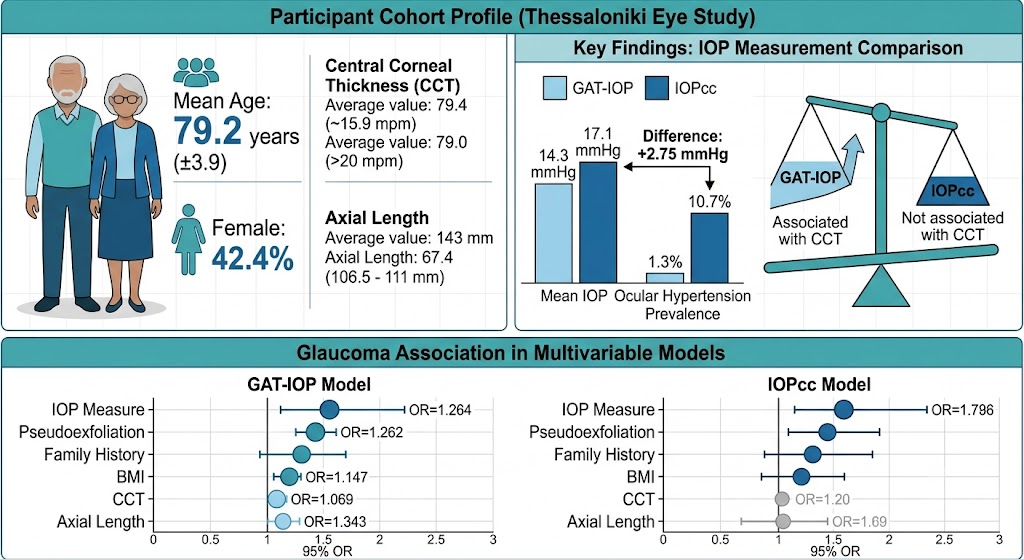
In this population-based study of 854 elderly participants, the Ocular Response Analyzer (IOPcc) produced systematically higher IOP readings than Goldmann applanation tonometry (GAT-IOP), with a mean difference of 2.75 mmHg and wide limits of agreement (−3.2 to 8.7 mmHg).
Switching from GAT-IOP to IOPcc increased the prevalence of ocular hypertension (IOP >21 mmHg) from 1.3% to 10.7%, with lower specificity (88.3% vs 98.6%) but higher sensitivity (44.7% vs 34.2%) for glaucoma detection.
Areas under the curve for glaucoma detection were similar between the two methods (0.818 vs 0.780; P=0.31).
In multivariable models, GAT-IOP (OR=1.264) showed a stronger independent association with glaucoma than IOPcc (OR=1.176); pseudoexfoliation, family history, and body mass index were significant in both models.
The two IOP measures are not interchangeable; using IOPcc with standard thresholds may substantially overestimate ocular hypertension prevalence and alter risk estimates.
Study Snapshot
Design: Prospective diagnostic accuracy and agreement analysis embedded in a population-based cohort (Thessaloniki Eye Study 12-year incidence phase).
Participants: 854 participants (one eye each), mean age 79.2 years (SD 3.9), 42.4% female.
Measurements: Goldmann applanation tonometry (GAT-IOP) and corneal-compensated IOP (IOPcc) by Ocular Response Analyzer.
Key Results: Mean GAT-IOP 14.3 mmHg, IOPcc 17.1 mmHg. Prevalence of IOP >21 mmHg: GAT 1.3%, IOPcc 10.7%. Sensitivity for glaucoma: GAT 34.2%, IOPcc 44.7%; Specificity: GAT 98.6%, IOPcc 88.3%; AUC: 0.818 vs 0.780 (P=0.31). Multivariable OR for GAT-IOP 1.264 (95% CI 1.182–1.359), for IOPcc 1.176 (95% CI 1.116–1.241).
Conclusion: IOPcc and GAT-IOP are not interchangeable; substituting may overestimate ocular hypertension and change risk models.
Why This Study Matters
Accurate intraocular pressure (IOP) measurement is central to glaucoma diagnosis and management. Goldmann applanation tonometry (GAT) has been the clinical standard for decades, but its readings are influenced by corneal thickness and other biomechanical properties. The Ocular Response Analyzer (ORA) provides a corneal-compensated IOP (IOPcc) that attempts to correct for these factors. However, the clinical impact of switching from GAT-IOP to IOPcc in a real-world, population-based setting—especially among older adults where glaucoma prevalence is highest—remains incompletely understood. The Thessaloniki Eye Study directly compared these two methods in a large, elderly cohort and examined how substituting one for the other would alter IOP distribution, ocular hypertension prevalence, and glaucoma screening performance.
How the Study Was Conducted
This investigation was part of the 12-year incidence phase of the Thessaloniki Eye Study, a population-based prospective cohort in Greece. Researchers enrolled 854 participants (one eye per person) who underwent both GAT and ORA measurements in the same session. The mean age was 79.2 years (SD 3.9); 42.4% were female. Glaucoma was defined by standard optic nerve and visual field criteria. Agreement between GAT-IOP and IOPcc was assessed using Bland-Altman plots and paired t-tests. Diagnostic performance for detecting glaucoma was evaluated with sensitivity, specificity, and area under the receiver operating characteristic curve (AUC). Two multivariable logistic regression models—one including GAT-IOP and the other IOPcc—were constructed to compare risk factor associations, adjusting for confounders such as age, sex, central corneal thickness (CCT), axial length, pseudoexfoliation, family history of glaucoma, and body mass index.
What the Researchers Found
The mean GAT-IOP was 14.3 mmHg (SD 3.3), while mean IOPcc was significantly higher at 17.1 mmHg (SD 4.3). The average difference (IOPcc minus GAT-IOP) was 2.75 mmHg, with 95% limits of agreement ranging from −3.2 to 8.7 mmHg, indicating poor agreement between the two methods. IOPcc was not associated with CCT (95% CI −0.001 to 0.016; P=0.08), whereas GAT-IOP remained significantly associated with CCT (95% CI 0.009–0.022; P=0.02), as expected.
Using a conventional threshold of IOP >21 mmHg, ocular hypertension prevalence rose from 1.3% with GAT to 10.7% with IOPcc—an eightfold increase. In untreated participants, GAT-IOP >21 mmHg had 98.6% specificity and 34.2% sensitivity for glaucoma detection; IOPcc >21 mmHg improved sensitivity to 44.7% but reduced specificity to 88.3%. The AUCs for the two methods were not significantly different (0.818 vs 0.780; P=0.31).
In the regression models, both IOP measures were independently associated with glaucoma, but the effect size for GAT-IOP (OR=1.264; 95% CI 1.182–1.359) was larger than that for IOPcc (OR=1.176; 95% CI 1.116–1.241). Pseudoexfoliation, family history of glaucoma, and body mass index were significant in both models. Central corneal thickness and axial length were significant only in the GAT-IOP model, suggesting that IOPcc partially accounts for corneal properties but may also introduce other sources of variation.
What the Findings May Mean
These results indicate that GAT-IOP and IOPcc are not interchangeable in an elderly population. The systematic upward shift of IOPcc values and the resulting spike in ocular hypertension prevalence—from 1.3% to 10.7%—raises concern that using IOPcc with the same threshold (>21 mmHg) could lead to substantial overdiagnosis of ocular hypertension and potentially unnecessary treatment or monitoring. The trade-off in screening performance (higher sensitivity, lower specificity) may be acceptable in certain research contexts but would likely increase false-positive referrals in clinical practice. The stronger association of GAT-IOP with glaucoma in the multivariable models also suggests that GAT-IOP may retain better prognostic value, perhaps because it reflects a longer history of clinical use and validated risk algorithms.
Strengths and Limitations
The study's primary strengths include its population-based design, large sample size for an elderly cohort, and direct head-to-head comparison of the two tonometry methods within the same individuals. The use of standardized glaucoma definitions and adjustment for multiple confounders also enhances the reliability of the findings. However, several limitations should be considered. This analysis is based solely on the published abstract; full details of the study protocol, exclusion criteria, masking procedures, and statistical methods were not available. The cross-sectional nature of the diagnostic accuracy comparison within a longitudinal cohort means that temporal relationships between IOP and glaucoma progression cannot be addressed. The participants are all of Greek ancestry, limiting generalizability to other ethnic groups. Additionally, the study used only one ORA device per patient and did not assess intra-session reproducibility of IOPcc. Finally, the optimal threshold for IOPcc in glaucoma screening was not determined; the analysis used the conventional >21 mmHg cutpoint for both methods, which may not be appropriate for IOPcc.
Implications for Practice and Research
Clinicians should be aware that ORA-derived IOPcc values are not equivalent to GAT-IOP and should not be interpreted using the same thresholds. If IOPcc is used, practice-specific reference ranges and decision points need to be established. For researchers, these findings highlight the importance of specifying the tonometry method in IOP-related analyses and cautions against interchangeable use in risk prediction models. Future studies should explore whether IOPcc-based definitions of ocular hypertension better predict glaucoma incidence or progression over time and whether device-specific thresholds could improve screening accuracy. Validation in other populations and with different ORA software versions is also warranted.
Funding, Disclosures, and Registration
Not reported in the abstract. The Thessaloniki Eye Study is a long-standing population-based project; institutional support and grant information are typically detailed in the full manuscript.
References
Bartzoulianou RC, Coleman AL, Wilson MR, et al. Ocular Response Analyzer versus Goldmann Applanation Tonometry in a Population-based Sample: The Thessaloniki Eye Study. American Journal of Ophthalmology. 2026; PMID: 42508710. https://pubmed.ncbi.nlm.nih.gov/42508710/
This article was created using several editorial tools, including AI, as part of the process. Human editors reviewed this content before publication.