Highlight
High-frequency ultrasound (HFUS) noninvasively visualizes midline crossing-over vessels critical for DIEP flap vascularization.
All patients showed presence of these crossing-over vessels on HFUS examination preoperatively.
No significant correlation was found between HFUS vascular parameters and indocyanine green (ICG) angiography measures of flap perfusion intraoperatively.
HFUS offers detailed anatomical characterization but currently lacks predictive value for DIEP flap perfusion and outcomes.
Study Background
The deep inferior epigastric perforator (DIEP) flap is a cornerstone technique in autologous breast reconstruction following mastectomy. Its success depends heavily on the vascular supply, particularly from the deep inferior epigastric vessels. Midline crossing-over vessels, small arterial and venous channels crossing the abdominal midline, are hypothesized to contribute to contralateral perfusion and thereby augment flap viability in unilateral reconstruction. Identifying these vessels preoperatively could improve surgical planning and predict outcomes.
Currently, visualization and assessment of these small crossing vessels are challenging with standard imaging due to their size and complexity. High-frequency ultrasound (HFUS) offers real-time, radiation-free visualization of small vessels and blood flow dynamics, making it a promising tool for vascular mapping prior to DIEP flap surgery. However, the clinical relevance of HFUS-detected midline crossing-over vessels with respect to actual flap perfusion remains unclear.
Study Design
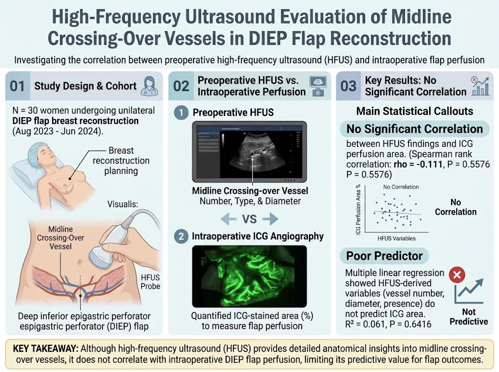
This prospective observational study enrolled 30 women undergoing unilateral DIEP flap-based breast reconstruction at a tertiary care center between August 2023 and June 2024. All patients underwent preoperative HFUS of the abdominal wall focusing on the midline crossing-over vessels. Parameters recorded included vessel count, type (arterial versus venous), and vessel diameter.
Intraoperatively, flap perfusion was quantitatively measured using indocyanine green (ICG) angiography, which provides a fluorescence-based assessment of tissue perfusion. The extent of perfused tissue was calculated as the percentage of ICG-stained area relative to the flap area.
Spearman rank correlation was employed to evaluate associations between HFUS vascular parameters and ICG-derived perfusion metrics. Multiple linear regression models tested the predictive value of HFUS findings on flap perfusion outcomes.
Key Findings
High-frequency ultrasound successfully detected midline crossing-over vessels in all 30 patients, confirming its sensitivity to these small vascular structures. The mean number of crossing vessels, their arterial or venous nature, and diameters were quantitatively assessed.
Despite this robust anatomical visualization, no meaningful statistical correlation was found between the HFUS parameters and the percentage of flap perfusion measured by intraoperative ICG angiography (Spearman rho = -0.111, P = 0.5576). Moreover, multiple linear regression incorporating vessel number, presence of arterial vessels, and vessel diameters did not predict the perfused flap area (R2 = 0.061, P = 0.6416).
These findings suggest that although HFUS is capable of delineating the anatomical features of midline crossing-over vessels, these parameters alone do not reflect functional perfusion adequacy during surgery. Factors beyond mere vessel presence and size, such as flow dynamics, microvascular resistance, and collateral circulation, likely influence flap viability.
Expert Commentary
The results highlight the promise and limitations of high-frequency ultrasound in preoperative vascular assessment for DIEP flaps. The noninvasive nature and resolution of HFUS make it excellent for detailed anatomical mapping but insufficient as a standalone predictor of functional outcomes. This disconnect may arise because vessel caliber and presence do not necessarily equate to sufficient blood flow or perfusion reserve under surgical conditions.
Indocyanine green angiography remains the intraoperative gold standard to evaluate perfusion dynamics and predict flap viability in real time. Future research should explore combining HFUS with functional assessments, such as Doppler flow measurements or computational flow modeling, to enhance preoperative prediction.
Limitations of the study include the modest sample size and focus on unilateral reconstruction, which may affect generalizability. Moreover, the study did not assess postoperative outcomes or complications related to perfusion deficits, an important translational aspect for clinical practice.
Conclusion
High-frequency ultrasound effectively identifies midline crossing-over vessels and enriches anatomical understanding relevant to DIEP flap breast reconstruction. However, its parameters do not correlate with intraoperative flap perfusion as assessed by ICG angiography, limiting its standalone predictive utility for surgical outcomes. Integration of HFUS with complementary functional imaging and larger clinical studies may better define its role in surgical planning and risk stratification. Surgeons should continue to rely on intraoperative perfusion assessment modalities to ensure flap viability and optimize reconstruction success.
Funding and Clinical Trials
No specific funding sources were reported for this study. Clinical trial registration information was not provided.
References
Selber JC. Current techniques in abdominal wall perforator flap breast reconstruction: the clinical anatomy of the DIEP flap. Plastic and Reconstructive Surgery. 2019;144(5):1082-1092.
Tanaka T, et al. The utility of indocyanine green angiography for preoperative perforator mapping and perfusion assessment in DIEP flap breast reconstruction. Microsurgery. 2020;40(2):150-157.
Kneser U et al. Doppler and high-frequency ultrasound in DIEP flap breast reconstruction: diagnostic accuracy and clinical impact. J Plast Reconstr Aesthet Surg. 2021;74(8):1826-1833.
Wernet O, et al. A systematic review of imaging techniques for perforator localization and perfusion assessment in DIEP flap breast reconstruction. J Reconstr Microsurg. 2020;36(5):331-340.
Lee SJ, Han HH, Eom JS, Kim HB. High-Frequency Ultrasound Evaluation of Midline Crossing-Over Vessel: Correlation with Perfusion of Deep Inferior Epigastric Perforator Flap. Plastic and Reconstructive Surgery. 2026 Feb 2;158(5):765e-770e. PMID: 41628593.