Evaluating CT-Based Models to Predict Residual Disease and Surgical Extent in Interval Debulking Surgery for Ovarian Cancer
Highlight
- Complete gross resection (CGR) is critical for survival in advanced ovarian cancer after neoadjuvant chemotherapy.
- The Memorial Sloan Kettering Cancer Center (MSKCC) CT-based model shows limited accuracy when applied to interval debulking surgery (IDS).
- Incorporating patient age, CA-125 biomarker thresholds, and selected radiologic features improves model prediction of residual disease and surgical complexity.
- Specific CT findings may suggest the need for advanced surgical techniques, but absence of such findings does not exclude the requirement for complex surgery.
Study Background
Advanced ovarian cancer (AOC), classified primarily as International Federation of Gynecology and Obstetrics (FIGO) stage IIIC-IV, carries a poor prognosis largely dictated by the extent of residual disease (RD) following cytoreductive surgery. Achieving complete gross resection (CGR) consistently correlates with improved survival. Treatment typically involves either primary debulking surgery or neoadjuvant chemotherapy (NACT) followed by interval debulking surgery (IDS). While predictive modeling based on preoperative computed tomography (CT) scans has shown promise in predicting RD and operative complexity in upfront surgery, its utility after NACT remains uncertain. Post-NACT tumor responses may alter disease distribution and imaging characteristics, which potentially limit existing model accuracy. Better tools to anticipate RD and surgical complexity post-NACT are needed to optimize surgical planning and patient selection for IDS.
Study Design
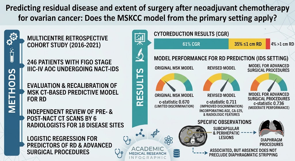
This was a multicenter retrospective cohort study conducted between 2016 and 2021, including 246 patients with FIGO stage IIIC-IV AOC undergoing NACT followed by IDS. The core objective was to evaluate the performance of the Memorial Sloan Kettering Cancer Center (MSKCC) CT-based predictive model—previously validated in primary surgery settings—to predict residual disease and surgical complexity after NACT. Both pre- and post-NACT CT scans were independently reviewed by six radiologists for 18 predefined abdominopelvic disease sites. Logistic regression analyses were employed to identify clinical and radiologic predictors of RD and the need for advanced surgical procedures such as diaphragmatic or perihepatic surgery. The MSK model was recalibrated using optimized age and CA-125 threshold biomarker levels to develop revised predictive models.
Key Findings
Among the cohort, complete gross resection (no visible RD) was achieved in 61% of patients, while 35% had residual disease ≤1 cm and 4% had residual disease >1 cm. When applying the existing MSK model to post-NACT IDS, discrimination for predicting RD was limited, with a c-statistic of 0.670, indicating modest performance. The authors developed a revised model that incorporated optimized thresholds for patient age and CA-125 biomarker levels along with selected radiologic features, improving the c-statistic to 0.711, reflecting enhanced predictive ability.
A separate logistic regression model aimed at predicting the requirement for advanced surgical procedures demonstrated moderate discrimination with a c-statistic of 0.736. Notably, lesions involving subcapsular liver and perihepatic sites on CT scans correlated with diaphragmatic surgeries. However, the absence of such lesions on imaging did not reliably exclude the necessity for diaphragmatic stripping or other complex procedures, underscoring limitations in sensitivity.
The data highlight that radiologic parameters alone are not entirely sufficient for surgical planning post-NACT. The integration of clinical markers like CA-125 and patient age improves predictions, reflecting the multifactorial nature of surgical complexity after chemotherapy-induced tumor alteration.
Expert Commentary
The study addresses a critical gap in ovarian cancer management by validating a widely referenced CT-based predictive tool in the NACT-IDS setting. The modest c-statistics reflect inherent challenges in imaging-based prediction post-chemotherapy, which alters tumor burden and tissue characteristics, potentially masking residual disease. This emphasizes the necessity of combining imaging with clinical biomarkers and multidisciplinary clinical judgment to guide surgical planning effectively.
Limitations of the study include its retrospective design and potential interobserver variability among radiologists, despite independent reviews. Furthermore, surgical decision-making encompasses factors beyond imaging and CA-125, such as patient performance status and intraoperative findings, which are difficult to model. This study reinforces that while models like MSK can assist in risk stratification and operative planning, a negative scan does not obviate the need for experienced surgical teams prepared for comprehensive cytoreduction.
These findings align with current NCCN guidelines emphasizing individualized surgical approaches in IDS and the value of biomarker integration in patient stratification. Future prospective studies should aim to refine predictive models by incorporating advanced imaging techniques, molecular biomarkers, and real-time surgical variables, potentially leveraging machine learning approaches to enhance prediction accuracy.
Conclusion
In conclusion, the MSKCC CT-based model, originally developed for primary ovarian cancer surgery, demonstrates only limited accuracy in predicting residual disease after neoadjuvant chemotherapy during interval debulking surgery. Recalibration of the model with clinical parameters such as age and CA-125 slightly improves discrimination but does not fully resolve predictive limitations. Specific CT findings are informative for anticipating complex surgical maneuvers, yet surgical planning must consider that the absence of radiologic evidence of disease does not preclude extensive operative intervention. Multidisciplinary assessment integrating clinical, radiologic, and biomarker data remains essential for optimizing surgical outcomes in advanced ovarian cancer post-NACT. This study underscores the need for enhanced predictive tools tailored to the post-chemotherapy surgical landscape to better guide patient selection and operative strategy.
Funding and Clinical Trials
The study does not report specific funding sources or clinical trial registration numbers. The retrospective nature and multicenter participation highlight collaborative efforts across institutions specializing in gynecologic oncology.
References
1. Ainio C, Caruso G, Alessi S, et al. Predicting residual disease and extent of surgery after neoadjuvant chemotherapy for ovarian cancer: Does the MSKCC model from the primary setting apply? Gynecologic Oncology. 2026 Aug 18;212:145-151. PMID: 42612479.
2. National Comprehensive Cancer Network. NCCN Clinical Practice Guidelines in Oncology: Ovarian Cancer Including Fallopian Tube Cancer and Primary Peritoneal Cancer. Version 1.2024.
3. Eisenhauer EA, et al. New response evaluation criteria in solid tumours: Revised RECIST guideline (version 1.1). Eur J Cancer. 2009 Jan;45(2):228-47.
4. Fagotti A, et al. Prospective validation study of a laparoscopic predictive model for optimal cytoreduction in advanced ovarian carcinoma. Am J Obstet Gynecol. 2008;199(6):617.e1-617.e6.
5. Rutten MJ, et al. Predictive diagnostic models of residual disease after primary debulking surgery in advanced-stage ovarian cancer. J Clin Oncol. 2019;37(25):2259-2269.
This article was created using several editorial tools, including AI, as part of the process. Human editors reviewed this content before publication.