Novel Peripheral Retinal Vascular Biomarker on Ultra-Widefield OCTA Enables Severity Grading in Retinitis Pigmentosa
peripheral retinal vasculatureultra-widefield OCT angiographydisease severity gradingretinitis pigmentosa
Highlight
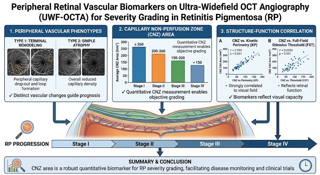
- Identification of two distinct peripheral retinal vascular phenotypes in retinitis pigmentosa (RP) via ultra-widefield OCT angiography (UWF-OCTA): Terminal Remodeling and Simple Atrophy.
- Validation of the chorioretinal neovascular zone (CNZ) area as a robust quantitative biomarker correlating strongly with multiple visual function measures.
- Development and validation of a clinically applicable four-stage severity grading system based on CNZ area reliably stratifying RP disease progression.
- Potential for the grading system to enhance disease monitoring and patient stratification in future interventional trials.
Study Background
MedXY registered readers
Sign in free to continue reading
Create or use your MedXY account to unlock the complete article.
This article was created using several editorial tools, including AI, as part of the process. Human editors reviewed this content before publication.