Insights into Postpartum Levator Ani Muscle Remodeling: Impact of Maternal Age and Recovery Duration on Pelvic Floor Health
Highlight
- Maternal age influences transcriptional remodeling in the levator ani muscle after first vaginal delivery, potentially affecting pelvic floor recovery.
- Age and recovery time modulate gene expression related to tissue repair, senescence, and p53-regulated checkpoints, implicating disrupted healing mechanisms.
- Transcriptomic signatures correlate strongly with clinical pelvic floor function, capturing biological changes beyond chronological age alone.
- Findings support mechanistic pathways for increased pelvic organ prolapse risk in older primiparas and suggest potential for targeted therapeutic interventions.
Study Background and Clinical Context
Pelvic organ prolapse (POP) is a prevalent condition characterized by descent of pelvic organs, often resulting in discomfort, urinary or fecal dysfunction, and diminished quality of life. Vaginal delivery is a well-established risk factor due to trauma sustained by the pelvic floor musculature, especially the levator ani muscle. Observational data have indicated that older maternal age at first vaginal delivery—commonly defined as 30 years or older—is associated with a substantially elevated risk of POP later in life. The biological underpinnings of this association remain incompletely understood.
The levator ani muscle complex plays an essential role in pelvic organ support, and its postpartum recovery is critical for long-term pelvic floor integrity. Age-related declines in muscle regenerative capacity and altered molecular repair pathways may impair healing, predisposing older women to dysfunction and prolapse. However, direct molecular data on levator ani remodeling after vaginal birth, stratified by maternal age, are scarce.
Study Design
This prospective, translational study enrolled nulliparous women in their third trimester, stratified into two groups by age: young primiparas aged 18-25 years, and older primiparas aged 35 years or older. Levator ani muscle biopsies were obtained immediately after vaginal delivery and again at 6 to 8 weeks postpartum. This paired sampling design enabled investigation of transcriptional changes associated with both delivery and subsequent recovery in the same individuals.
At the postpartum visit, clinical assessments included measures of pelvic floor support and levator ani muscle function to establish phenotypic correlations. High-throughput RNA sequencing analyses were performed on biopsy samples to identify differentially expressed genes (DEGs) by maternal age, time point, and their interaction. Statistical rigor was ensured using moderated t-tests controlling false discovery rate below 5%, and principal component analysis (PCA) was employed to elucidate relationships between transcriptomic profiles and clinical phenotypes.
Key Findings
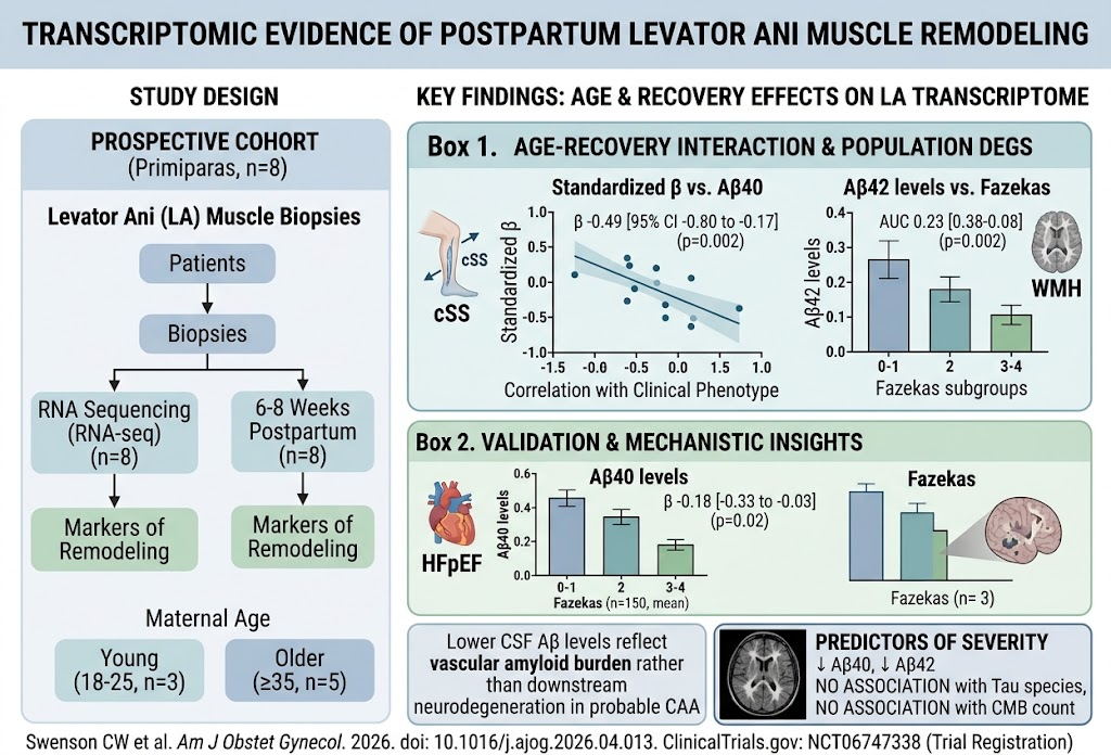
A total of eight women (three young and five older) provided sufficient biopsies at both time points for transcriptomic analyses.
1. **Age and Recovery Interaction:** Fifty-eight genes were differentially expressed when considering the interaction of maternal age and recovery interval. Notably, elevated expression of TP53BP1, PRR12, and ZNF316 and reduced expression of PLK3 were identified. TP53BP1 encodes a critical DNA damage response protein regulating p53 checkpoints, while PLK3 is involved in cell cycle regulation and senescence pathways.
2. **Age-Associated Differences:** Comparing age groups alone yielded 309 DEGs, highlighting pronounced age-related shifts in musculature remodeling gene networks.
3. **Time-Dependent Differences:** Comparing early postpartum biopsies to those obtained after 6 to 8 weeks identified 159 DEGs, consistent with ongoing healing and tissue repair processes.
4. **Biological Pathways:** Both age groups demonstrated enrichment in core tissue repair and homeostasis pathways, yet the gene expression patterns suggest that aging may alter the balance between effective repair and cellular senescence.
5. **Correlations with POP Studies:** Twenty genes identified in age- or time-dependent analyses overlapped with previously reported genes from case-control studies of pelvic organ prolapse, reinforcing the clinical relevance of the findings.
6. **Transcriptomic-Clinical Correlation:** PCA revealed a robust positive correlation (ρ=0.88, false discovery rate <0.05) between principal components representing transcriptomic signatures and clinical pelvic floor function, whereas correlation with chronological maternal age alone was weaker and inverse (ρ=-0.72). This indicates that gene expression patterns more accurately reflect pelvic floor condition than age per se.
Expert Commentary and Mechanistic Insights
This investigation breaks new ground by linking postpartum levator ani muscle molecular remodeling with maternal age and recovery time. The observed differential expression of TP53BP1 and PLK3 implicates dysregulation of p53-dependent DNA repair and checkpoint pathways, which are pivotal for cellular integrity and regeneration. Elevated TP53BP1 combined with reduced PLK3 suggests a phenotype characterized by heightened cellular senescence— a state where cells cease proliferation and repair, contributing to dysfunctional tissue remodeling.
The study’s integration of transcriptomic data with clinical outcomes highlights that biological aging in muscle tissue extends beyond chronological age, possibly influenced by individual variability in regenerative capacities or environmental factors.
While the sample size is limited and further validation is warranted, this work supports a model wherein the levator ani of older primiparas demonstrates an altered recovery trajectory at the molecular level, which may underlie their increased vulnerability to pelvic organ prolapse. Understanding these pathways could facilitate development of targeted therapies—pharmacological agents or regenerative strategies—that promote more favorable recovery after vaginal delivery.
Limitations and Future Directions
The relatively small cohort and single-center design limit broad generalizability. Larger studies encompassing diverse populations and extending follow-up beyond the early postpartum period would clarify the durability and functional significance of the transcriptional changes. Furthermore, elucidating the impact of other factors such as parity, delivery trauma severity, and comorbidities will refine risk stratification. Functional studies validating the roles of candidate genes like TP53BP1 and PLK3 in levator ani biology are also warranted.
Conclusion
By revealing distinct transcriptional signatures associated with maternal age and postpartum recovery in the levator ani muscle, this study sheds light on the molecular basis for impaired pelvic floor healing and increased pelvic organ prolapse risk in older first-time mothers. These findings represent a significant advance toward personalized postpartum care and potential preventative interventions, emphasizing the importance of considering biological remodeling processes when assessing pelvic floor health after vaginal delivery.
Funding and Registration
Details of funding sources and clinical trial registration were not provided in the source abstract.
References
Swenson CW, Pouladi N, Zabriskie HA, Bourrant PE, Li J, Wilson LS, Drummond MJ, Lussier YA. Transcriptomic evidence of remodeling in postpartum levator ani muscle associated with maternal age and recovery time after first vaginal delivery. Am J Obstet Gynecol. 2026 Apr 15;235(3):594-607. PMID: 41997518.
Additional supporting literature:
– Bradley CS, Nygaard IE. Pelvic organ prolapse and aging. Obstet Gynecol Clin North Am. 2005;32(3):481-497.
– Kandadai MR, et al. Age-related changes in the connective tissue and muscle of the levator ani. Neurourol Urodyn. 2013;32(4):316-320.
This article was created using several editorial tools, including AI, as part of the process. Human editors reviewed this content before publication.