Normal Late-Third Trimester Ultrasound Does Not Rule Out Adverse Outcomes, Retrospective Cohort Study Finds
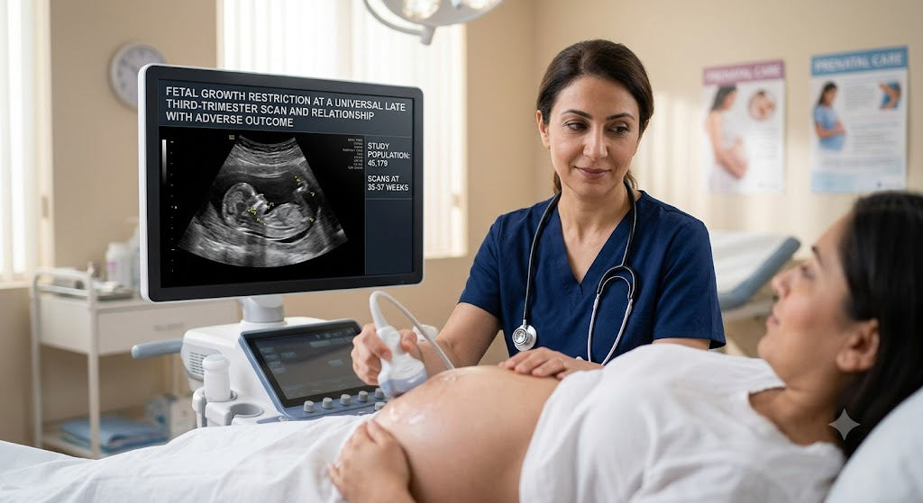
In a cohort of over 45,000 pregnancies, the absolute risk of stillbirth and composite adverse outcomes was similar among fetuses with fetal growth restriction (FGR) and those classified as normal on a routine late third-trimester scan.
More than 79% of stillbirths and 81% of composite adverse outcomes occurred in pregnancies with a normal scan, despite a 62.7% detection rate for severe small-for-gestational-age (SGA) at birth.
The findings highlight that a normal ultrasound does not guarantee a low-risk outcome and that intervention may mitigate risk in diagnosed FGR cases.
Study Snapshot
Design
Retrospective population-based cohort study
Setting
John Radcliffe Hospital, Oxford, UK (universal ultrasound at 35+1–36+6 weeks)
Population
45,179 singleton pregnancies without congenital abnormalities, excluding births before scan
Exposure
Five mutually exclusive phenotypes: ISUOG FGR (Delphi criteria), constitutional SGA (EFW <10th centile), AGA with abnormal Doppler (cerebroplacental ratio <5th centile or umbilical artery PI >95th centile), AGA with slow abdominal circumference growth velocity (ACGV <10th centile), or normal AGA
Primary Outcomes
Stillbirth, composite adverse outcome (Grade 2–3 encephalopathy, cooling, ventilation >24 h, or perinatal death), severe SGA at birth, neonatal unit admission, obstetric interventions
Main Finding
Absolute risk of stillbirth and composite adverse outcome was 0.1%–0.2% across all groups; the majority of adverse events occurred in the normal-AGA group (79.6% of stillbirths, 81% of composite outcomes).
Why This Study Matters
Fetal growth restriction (FGR) and small-for-gestational-age (SGA) are recognized risk factors for adverse perinatal outcomes, prompting many centers to adopt universal late third-trimester ultrasound screening. However, the ability of such screening to identify pregnancies at risk for stillbirth, neonatal morbidity, and other adverse outcomes remains debated. This large retrospective cohort study from a UK hospital where universal ultrasound is standard practice provides a detailed look at the association between different ultrasound phenotypes and outcomes near term.
How the Study Was Conducted
Researchers analyzed data from 45,179 singleton pregnancies undergoing a routine ultrasound scan between 35+1 and 36+6 weeks’ gestation at the John Radcliffe Hospital in Oxford, UK. Pregnancies with congenital abnormalities and those delivering before the scan were excluded. Using a hierarchical classification, they assigned each fetus to one of five mutually exclusive phenotypes: FGR by the International Society of Ultrasound in Obstetrics and Gynecology (ISUOG) Delphi consensus criteria; constitutional SGA (estimated fetal weight [EFW] <10th centile); appropriate-for-gestational-age (AGA) with abnormal cerebroplacental ratio (<5th centile) or umbilical artery pulsatility index >95th centile; AGA with slowing abdominal circumference growth velocity (ACGV <10th centile); or normal AGA (none of the above). The normal-AGA group served as the reference in univariate logistic regression analyses for stillbirth and a composite adverse outcome (CAO) defined as one or more of: Grade 2–3 encephalopathy, therapeutic cooling, mechanical ventilation >24 h, or perinatal death. Group differences in continuous variables were assessed using generalized linear models.
What the Researchers Found
Among the 45,179 pregnancies, 54 stillbirths (0.1%) and 253 composite adverse outcomes (0.6%) occurred. The normal-AGA group comprised 82% of the cohort and accounted for 43 stillbirths (79.6%) and 205 composite adverse outcomes (81.0%). In contrast, the FGR group represented only 1.9% of the cohort but had a similar absolute risk of stillbirth (0.2%) and composite adverse outcome (0.2%) as the normal-AGA group (0.1% and 0.2%, respectively). The constitutional SGA group and the two AGA-abnormal subgroups all showed similarly low absolute risks (0.1%–0.2%). The ultrasound screening detected 62.7% of neonates subsequently born with severe SGA (birth weight <3rd centile), yet most adverse outcomes originated from the large normal-AGA population.
What the Findings May Mean
The authors suggest that the similar perinatal risks across phenotypes likely reflect the effect of obstetric intervention, including early delivery, in pregnancies identified as FGR or SGA at the scan. In other words, detection and management of suspected growth restriction may mitigate the baseline risk, whereas the majority of adverse outcomes occur in the many pregnancies classified as normal and thus not considered high-risk. This underscores the limitations of a single late third-trimester scan as a standalone tool for predicting adverse outcomes: a “normal” result does not reliably exclude risk. The findings also highlight that even a relatively high detection rate for severe SGA does not translate into a proportional reduction in population-level adverse outcomes, because the vast majority of events arise from the large low-risk group.
Strengths and Limitations
Strengths of the study include its large, unselected population and the use of a universal screening protocol with standardized ultrasound phenotyping based on international criteria. However, several limitations must be considered. The retrospective, single-center design limits generalizability, and the observed outcomes may be influenced by local clinical management protocols and the threshold for intervention. The authors note that causality cannot be inferred due to the observational design, and unmeasured confounders—including details of obstetric management after the scan—may affect the associations. Additionally, the study period and specific ultrasound techniques may not reflect current practice in other settings.
Implications for Practice and Research
These results reinforce the importance of not overinterpreting a normal late third-trimester scan as low-risk. Clinicians should continue to consider other clinical risk factors, maternal symptoms, and serial growth assessment when making management decisions. For researchers, the findings point to the need for better risk-stratification tools that incorporate multiple parameters—including maternal characteristics, longitudinal growth patterns, and perhaps biomarkers—to identify the small number of pregnancies destined for adverse outcomes within the large normal-appearing group. Future prospective studies with standardized protocols and objective outcome ascertainment would help validate these observations.
References
D'Alberti E, Granieri C, Ioannou C, Aye CY, Shea M, Impey L. Fetal Growth Restriction at a Universal Late Third-Trimester Scan and Relationship With Adverse Outcome: Retrospective Cohort Study. BJOG. 2026;133(8):1592-1601. PMID: 41796020.