Evaluating the Safety of High-Grade Percutaneous MCL Pie-Crusting in Medial Meniscus Surgery: Implications for Valgus Stability
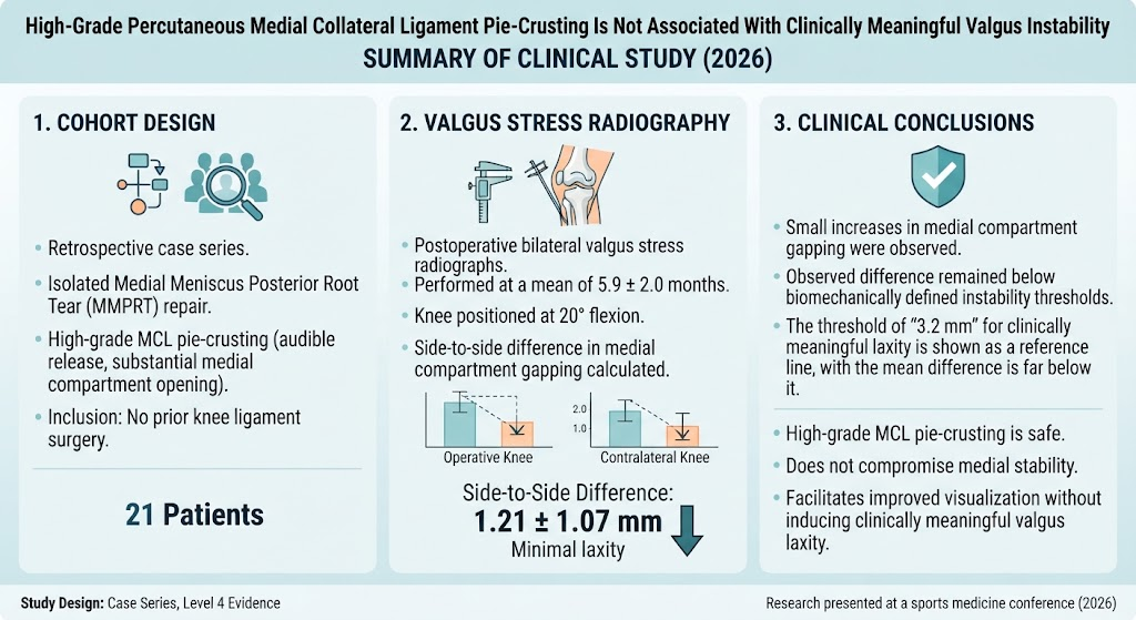
Highlight
High-grade percutaneous MCL pie-crusting significantly improves medial compartment visualization during medial meniscus posterior root tear (MMPRT) repair.
Postoperative valgus stress radiographs show only minor increases in medial compartment gapping after high-grade MCL pie-crusting.
The increase in medial laxity remains well below the biomechanically defined threshold for clinically meaningful valgus instability.
These results support the safety of an aggressive pie-crusting release technique without compromising knee stability.
Background
Medial meniscus posterior root tears (MMPRTs) are a significant cause of knee dysfunction and predispose to rapid degenerative changes within the joint. Surgical repair of MMPRT is often technically demanding due to limited medial compartment visualization and risk of iatrogenic cartilage injury. To address these challenges, percutaneous medial collateral ligament (MCL) pie-crusting has been employed intraoperatively to increase medial joint space and improve visualization.
Pie-crusting traditionally involves controlled incremental perforations of the MCL to release tension and widen the compartment. However, some surgeons extend these releases beyond routine incremental perforations, termed “high-grade” pie-crusting, which raises concerns regarding potential resultant valgus instability. The MCL is a primary restraint against valgus loads in the knee; hence, excessive release might hypothetically lead to medial laxity and clinical instability postoperatively.
Study Design and Methods
This study was a retrospective case series (Level of Evidence 4) reviewing patients who underwent isolated MMPRT repair combined with high-grade percutaneous MCL pie-crusting, excluding those with previous ligament surgeries on the knee.
High-grade pie-crusting was defined intraoperatively by the presence of both an audible and palpable release of the MCL and a substantial medial compartment opening of at least 6 mm under arthroscopic visualization at approximately 20° knee flexion.
Postoperative bilateral valgus stress radiographs were obtained at an average of 5.9 ± 2.0 months. Medial compartment gapping was quantitatively measured in millimeters for both operative and contralateral knees, with the contralateral knee serving as an internal control. Side-to-side differences were calculated. A prior power analysis determined that at least 15 patients were needed to detect a clinically meaningful difference defined as an increased gapping of 3.2 mm, consistent with established biomechanical instability thresholds.
A one-sample t-test compared the mean side-to-side difference to the 3.2-mm clinical cutoff.
Key Findings
A total of 21 patients fulfilled inclusion criteria. The operative knees showed a mean medial compartment gapping of 7.84 ± 1.65 mm, whereas the contralateral knees measured 6.62 ± 1.37 mm. This resulted in a mean side-to-side difference of 1.21 ± 1.07 mm (95% confidence interval [CI]: 0.73 to 1.70 mm), which was significantly less than the threshold of 3.2 mm for clinically meaningful valgus instability (P < .001).
No patients exhibited symptoms or clinical signs of valgus instability on postoperative evaluation. Furthermore, the degree of medial space opening intraoperatively correlated with improved visualization but did not translate into a destabilizing effect on the knee joint.
These findings suggest that the practice of extending the MCL release to a high-grade level is biomechanically safe in terms of valgus laxity.
Expert Commentary
The clinical utility of percutaneous MCL pie-crusting during medial meniscus surgery is well recognized for enhancing surgeon access and reducing iatrogenic cartilage damage. Prior biomechanical studies have established thresholds of medial laxity that correlate with symptomatic knee instability.
This study adds valuable clinical evidence by directly measuring valgus laxity radiographically and benchmarking against contralateral knees. The statistically and clinically insignificant increase in medial gapping supports the biological plausibility that the MCL has sufficient intrinsic healing capacity and residual ligamentous restraint post-pie-crusting.
Nevertheless, some limitations include retrospective design, the single surgical setting, and reliance on radiographic measures at a single time point postoperatively. Further prospective studies with longer follow-up and functional clinical assessment would be beneficial to confirm these results and explore patient-reported outcomes.
Conclusion
High-grade percutaneous MCL pie-crusting during MMPRT repair, defined by intraoperative audible release and substantial medial compartment opening, results in minor increases in medial compartment gapping that do not reach a level associated with clinical valgus instability.
This approach appears safe and effective for improving visualization and instrumentation during surgery without compromising postoperative knee stability. Surgeons may consider employing higher-grade pie-crusting techniques without undue concern for inducing valgus laxity, although careful intraoperative assessment and postoperative monitoring remain advisable.
Funding and ClinicalTrials.gov
The original study did not report specific funding sources or clinical trial registration identifiers.
References
Nishioka T, Thamrongskulsiri N, Casanova F, et al. High-Grade Percutaneous Medial Collateral Ligament Pie-Crusting Is Not Associated With Clinically Meaningful Valgus Instability. Am J Sports Med. 2026 Oct 12;3635465261492229. doi: 10.1177/03635465261492229. PMID: 42865127.
LaPrade RF, Wijdicks CA. Evaluation and treatment of medial collateral ligament injuries of the knee: an evidence-based review. Sports Med Arthrosc Rev. 2012 Mar;20(1):60-7. doi: 10.1097/JSA.0b013e318245d66b.
Dunn WR, Spindler KP. Surgical treatment of meniscal root injuries. Curr Opin Orthop. 2017;28(1):58-62. doi: 10.1097/BCO.0000000000000440.
This article was created using several editorial tools, including AI, as part of the process. Human editors reviewed this content before publication.