Highlight
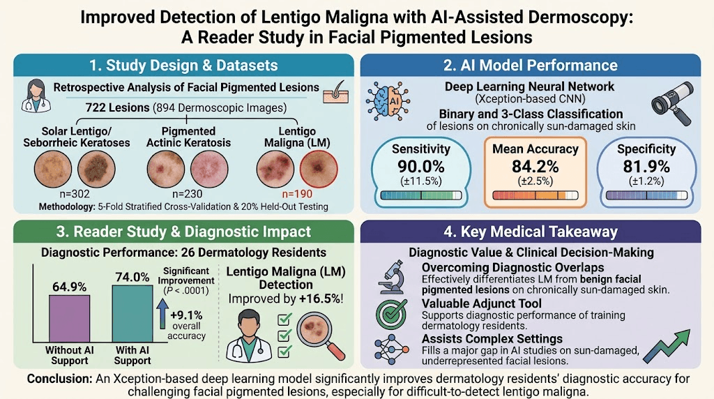
An AI model based on deep learning achieved 84.2% accuracy differentiating lentigo maligna (LM) from benign facial pigmented lesions.
AI assistance improved dermatology residents’ diagnostic accuracy from 64.9% to 74.0%, with a 16.5% gain specifically in LM detection.
The study focused on facial lesions in chronically sun-damaged skin, a challenging and underrepresented setting in AI skin lesion research.
Findings support AI as an adjunct tool for improving diagnostic confidence and educational value in clinical dermatology.
Study Background
Lentigo maligna (LM) is a melanoma in situ subtype occurring predominantly on chronically sun-exposed facial skin in elderly patients. Clinical and dermoscopic differentiation of LM from benign pigmented lesions—such as pigmented actinic keratosis, solar lentigo, or seborrheic keratosis—is notoriously challenging due to overlapping features, especially in sun-damaged skin. Accurate early detection is vital for appropriate management and prevention of progression to invasive melanoma. However, conventional diagnostic approaches may suffer from observer variability and require experienced dermatologists for expertise.
Artificial intelligence (AI), specifically convolutional neural networks (CNN), has shown promise for automated skin lesion classification. Nevertheless, previous AI models have underrepresented lesions on chronically sun-damaged facial skin, limiting their generalizability for LM diagnosis. This unmet diagnostic need for improved, accessible tools motivated this retrospective study to develop a deep learning model tailored to facial pigmented lesions and to evaluate its influence on dermatology resident diagnostic performance.
Study Design
This retrospective study analyzed 722 histopathologically confirmed facial pigmented lesions, comprising 190 LMs, 230 pigmented actinic keratoses (PAK), and 302 solar lentigines/seborrheic keratoses (SL/SK). A total of 894 dermoscopic images were utilized. The dataset was split with 20% reserved as an independent test set and the remaining 80% used for five-fold stratified cross-validation.
The deep learning architecture used was an Xception-based convolutional neural network trained for both binary classification (LM vs non-LM) and three-class classification (LM, PAK, SL/SK). Subsequently, a reader study was conducted involving 26 dermatology residents. Residents were asked to diagnose the lesions first unaided and then with AI model assistance to assess the impact on diagnostic accuracy.
Key Findings
The AI model achieved a mean classification accuracy of 84.2% ± 2.5%, with a high sensitivity of 90.0% ± 11.5% and specificity of 81.9% ± 1.2% for LM detection. These metrics reflect robust performance in distinguishing LM from other facial pigmented lesions despite the complex background of sun damage.
In the reader study, dermatology residents’ baseline diagnostic accuracy was 64.9%. When provided with AI assistance, their accuracy significantly improved to 74.0% (p < .0001). Notably, the largest improvement was observed in diagnosing LM, with a 16.5% increase in accuracy, highlighting the AI tool’s clinical relevance for melanoma in situ detection.
The findings demonstrate that AI assistance can effectively augment human diagnostic capability, particularly for trainees, offering improved diagnostic confidence and potentially earlier detection of LM.
Expert Commentary
This study provides important translational insight into the utility of AI in a nuanced clinical challenge—distinguishing LM from benign pigmented lesions on photodamaged facial skin. The use of a well-curated dataset reflecting a high-risk patient subgroup adds to its clinical relevance.
Limitations include its retrospective design and focus on an image-only dataset without integrating clinical context (e.g., patient history, lesion evolution). Moreover, the reader study exclusively involved dermatology residents, so results may not generalize to community physicians or expert dermatologists. Prospective validation and inclusion of multimodal data could enhance model performance and applicability.
Recent guidelines emphasize the need for adjunctive tools in dermoscopy training and clinical decision support. AI’s demonstrated ability to increase resident diagnostic accuracy supports its integration into dermatology education and practice. However, human oversight remains paramount to account for atypical presentations and individual patient factors.
Conclusion
This study highlights the promise of AI-assisted dermoscopy for improving the detection accuracy of lentigo maligna amidst challenging facial pigmented lesions on sun-damaged skin. The significant improvement in dermatology resident diagnostic performance indicates that such AI tools may serve as valuable adjuncts in both clinical practice and dermatology training.
Future research should focus on prospective, real-world implementation studies, integration with clinical metadata, and evaluation across varied user expertise levels to validate and optimize AI models. Ultimately, combining AI with clinician judgment offers a practical avenue to enhance early LM diagnosis and potentially patient outcomes.
Funding and ClinicalTrials.gov
The original study did not report specific funding sources or ClinicalTrials.gov registration, consistent with its retrospective design.
References
1. Yilmaz A, Erol Mart HM, Temelkuran B, Akay BN. Improved detection of lentigo maligna with AI-assisted dermoscopy: A reader study in facial pigmented lesions. J Am Acad Dermatol. 2026;95(4):1035-1042. PMID: 42336217.
2. Ferris LK, Wiggins T, Schmults CD. Dermatologic Surgery in High-Risk Patients: Approaches to Lentigo Maligna. Dermatol Clin. 2021;39(3):341-350.
3. Esteva A, Robicquet A, Ramsundar B, et al. A guide to deep learning in healthcare. Nat Med. 2019;25(1):24-29.
4. Kittler H, Pehamberger H, Wolff K, Binder M. Diagnostic accuracy of dermoscopy. Lancet Oncol. 2002;3(3):159-165.
5. Tschandl P, Rinner C, Apalla Z, et al. Human–computer collaboration for skin cancer recognition. Nat Med. 2020;26(8):1229-1234.