Differential Impact of Reticular Pseudodrusen Subtypes on Retinal Sensitivity in Intermediate Age-Related Macular Degeneration
Highlight
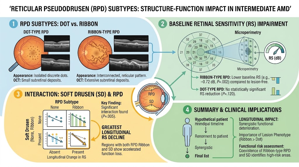
Ribbon-type reticular pseudodrusen (RPD) are associated with significantly lower retinal sensitivity (RS) at baseline than dot-type RPD or lesion-free regions in intermediate age-related macular degeneration (AMD).
Soft drusen (SD) independently reduce retinal sensitivity, and their coexistence with ribbon-type RPD results in the greatest functional decline over time.
No significant baseline interaction between RPD subtypes and SD was observed, but longitudinal analysis revealed synergistic detrimental effects on RS decline when both lesions coexist.
Lesion phenotype specificity is crucial for accurate functional risk stratification in intermediate AMD.
Study Background
Age-related macular degeneration (AMD) is a leading cause of irreversible visual impairment among the elderly worldwide. Intermediate AMD is marked by the presence of distinctive extracellular deposits beneath the retina, chiefly soft drusen (SD) and reticular pseudodrusen (RPD), which are implicated in disease progression to late AMD forms causing vision loss. RPD, also called subretinal drusenoid deposits, have been recognized as important risk factors for AMD progression. Recent advances have identified distinct RPD subtypes—primarily dot and ribbon forms—differing in morphology and possibly pathogenic mechanisms. However, their respective impacts on retinal function, alone or in combination with SD, have been incompletely characterized.
Understanding the interaction of RPD subtypes with SD on retinal sensitivity (RS) is pivotal. RS, assessed via microperimetry, offers a sensitive functional measure correlating with structural changes in the macula. A nuanced evaluation of how distinct RPD phenotypes influence RS over time, and how their coexistence with SD compounds the functional deficit, could inform more precise prognostic assessments and tailored clinical management strategies for intermediate AMD patients.
Study Design
This retrospective cohort study analyzed 46 eyes from 28 patients diagnosed with intermediate AMD or exhibiting RPD, studied longitudinally over 6 to 12 months. All participants underwent detailed microperimetry assessments at two time points, enabling the measurement of pointwise retinal sensitivity changes.
Ocular imaging included multimodal techniques to identify and subtype RPD lesions into dot and ribbon patterns, and to detect the presence of soft drusen. These imaging findings were localized to 37 individual test points aligned with the Early Treatment Diabetic Retinopathy Study (ETDRS) grid, permitting detailed spatial correlation between morphology and function.
Linear mixed-effects models were employed to evaluate baseline retinal sensitivity differences and longitudinal changes according to drusen composition at the test-point level. Models adjusted for potential confounders including test-point location and baseline RS for the change analysis.
Key Findings
At baseline, regions containing ribbon-type RPD demonstrated significantly reduced retinal sensitivity compared to lesion-free areas (mean difference 0.72 ± 0.24 dB, P = .002). In contrast, dot-type RPD regions did not show a statistically significant difference from non-lesion areas (P = .120). Independently, soft drusen were also associated with decreased RS (P = .021).
No significant baseline interaction effect between RPD subtype and SD presence on RS was detected, indicating their independent impact at this time point.
Crucially, longitudinal analyses revealed a significant interaction (P = .005) between RPD subtype and SD. Test points with both ribbon-type RPD and soft drusen showed the greatest decline in RS over the follow-up period. In contrast, RPD subtypes displayed no significant difference in RS progression when soft drusen were absent (all P > .9).
These results indicate that while the ribbon-type RPD exerts an inherent detrimental effect on retinal function, this is substantially exacerbated when soft drusen coexist, highlighting a compounded risk for progressive functional loss.
Expert Commentary
This study advances our understanding of AMD pathophysiology by dissecting how specific RPD phenotypes differentially affect retinal function and interact with other established risk lesions such as soft drusen. The identification that ribbon-type RPD places patients at higher functional risk, especially in the presence of SD, suggests that combining lesion morphology with functional testing can refine risk stratification beyond traditional clinical grading.
Methodologically, leveraging co-registered multimodal imaging and microperimetry on a pointwise basis enabled high-resolution structure-function mapping, an approach that can help unravel complex lesion interplay in AMD. The use of linear mixed models accounting for spatial and baseline function variations strengthens the robustness of the findings.
Limitations include the retrospective design and relatively short follow-up, which may not capture longer-term progression dynamics or ultimate visual outcomes such as acuity decline. Additionally, the sample size limits subgroup analyses and generalizability. Future prospective studies with larger cohorts and multimodal functional assessments like adaptive optics microperimetry or electrophysiology could validate and extend these findings.
Biologically, ribbon-type RPD may reflect more extensive or active subretinal alterations disrupting photoreceptor health, which when combined with the metabolic burden of soft drusen aggregates could synergistically impair retinal sensitivity.
Conclusion
The functional impact of reticular pseudodrusen in intermediate AMD is subtype-specific, with ribbon-type lesions conferring greater impairment. This effect is markedly intensified when ribbon-type RPD coexist with soft drusen, resulting in accelerated retinal sensitivity decline. These findings emphasize the importance of detailed lesion phenotyping using multimodal imaging combined with functional testing for personalized prognostication and patient monitoring.
Recognizing lesion phenotype interactions will aid clinicians in identifying patients at highest risk for functional deterioration and may guide future therapeutic intervention targeting specific AMD subtypes to preserve vision.
Funding and Registration
The original article does not specify funding sources or clinical trial registration information.
References
1. Kim M, Park Y, Choi SW, et al. Reticular Pseudodrusen Subtypes and Their Interaction With Soft Drusen: Longitudinal Structure-Function Analysis. Am J Ophthalmol. 2026 Sep 7; PMID: 42705516.
2. Zweifel SA, Spaide RF, Curcio CA, et al. Reticular pseudodrusen are subretinal drusenoid deposits. Ophthalmology. 2010;117(2):303-312.
3. Spaide RF. Reticular pseudodrusen and age-related macular degeneration. Retina. 2013;33(2):301-316.
4. Ouyang Y, Heussen FM, Dubis AM, et al. Retinal sensitivity and microstructural changes associated with reticular pseudodrusen in age-related macular degeneration. Retina. 2019;39(10):2025-2033.
This article was created using several editorial tools, including AI, as part of the process. Human editors reviewed this content before publication.