Highlight
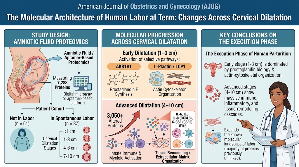
This comprehensive proteomic analysis defines the molecular architecture of the execution phase of human labor at term. Key highlights include: extensive proteomic remodeling during established labor; early labor (1–3 cm dilatation) marked by increased prostaglandin-related and cytoskeletal proteins; progressive expansion of molecular complexity with advancing cervical dilatation; and a dominant signature of immune activation and tissue remodeling processes at later stages. This study substantially expands the known molecular players involved in labor.
Study Background
Human parturition, or labor, is a complex physiological process involving coordinated biochemical and biophysical changes. It comprises a preparatory phase that primes the reproductive tissues, followed by the execution phase, characterized clinically by cervical dilatation and uterine contractions culminating in delivery. Despite the central role of labor in obstetric care, the molecular mechanisms governing its phases, especially the execution phase at term, remain incompletely understood. Understanding these mechanisms is key to informing interventions for labor-related complications such as preterm birth, labor dystocia, and cesarean delivery.
Amniotic fluid bathes the fetus and fetal membranes, reflecting the intrauterine environment’s molecular milieu. Proteomic profiling of amniotic fluid offers a window into the dynamic molecular events during labor. Prior studies have implicated inflammatory mediators and extracellular matrix remodeling proteins in labor; however, these studies have been limited by sample size, gestational windows, or breadth of proteomic coverage.
Study Design
This was a retrospective cross-sectional analysis involving 98 term pregnant women undergoing cesarean delivery with intact membranes. Participants were stratified based on labor status: 61 women not in labor and 37 women in spontaneous labor. Laboring women were further categorized by cervical dilatation into four groups: less than 1 cm, 1–3 cm, 4–6 cm, and 7–10 cm.
Amniotic fluid samples were collected intraoperatively, and an advanced aptamer-based proteomics platform quantified the abundance of 7,288 proteins, providing extensive proteomic coverage. The study assessed differential protein abundance comparing laboring versus non-laboring women and analyzed how protein expression patterns changed with increasing cervical dilatation. Functional enrichment analyses identified biological processes enriched within altered proteins.
Key Findings
1. Broad Proteomic Remodeling Associated with Labor
Established labor induced large-scale changes in the amniotic fluid proteome, with 656 proteins differentially abundant relative to non-laboring women. Among these, 214 proteins were increased, and 442 decreased, suggesting both induction and suppression of specific molecular pathways during labor.
2. Early Labor-Specific Molecular Changes (1–3 cm Dilatation)
At 1–3 cm cervical dilatation, distinct upregulation of aldo-keto reductase family 1 member B1 (AKR1B1) and L-plastin (LCP1) was observed. AKR1B1’s prostaglandin F synthase activity implicates prostaglandin signaling, a key mediator of uterine contractility and cervical ripening. LCP1, an actin-bundling protein, suggests cytoskeletal reorganization critical for cellular architecture change in the cervix and fetal membranes.
3. Progressive Molecular Complexity with Advancing Cervical Dilatation
The number of differentially abundant proteins increased markedly with cervical dilatation: 359 proteins at 4–6 cm and 3,050 proteins at 7–10 cm compared to non-laboring women. This indicates a substantial intensification and diversification of molecular processes as labor progresses to the active and transition phases.
4. Protein Association with Cervical Dilatation Within Laboring Women
Among laboring women, 580 proteins correlated with cervical dilatation, predominantly increasing with dilation (573 increased versus 7 decreased). Prominent inflammatory mediators such as interleukin-6 (IL-6), interleukin-8 (IL-8), granulocyte colony-stimulating factor (G-CSF), interferon gamma-inducible protein 16 (IFI16), neutrophil cytosol factor 1 (NCF1), and inducible T-cell costimulator ligand (ICOSL) were involved. This corroborates an immune-inflammatory milieu escalating with labor progression.
5. Functional Enrichment of Biological Processes
Proteins altered during labor were enriched in diverse biological processes: cell-to-cell communication, cell-junction and extracellular matrix (ECM) organization, tissue morphogenesis, cell adhesion, innate immune activation, and myeloid and neutrophil activation/degranulation. Cytokine signaling and RNA processing pathways were also identified, outlining a complex interplay of immune response, tissue remodeling, and cellular communication orchestrating cervical and uterine changes.
6. Expansion of Molecular Landscape of Human Labor
Remarkably, most proteins associated with labor in this study were novel in the context of parturition, significantly enlarging the catalog of molecular participants. This provides new avenues for biomarker discovery and mechanistic exploration.
Expert Commentary
This landmark proteomic study elucidates the dynamic molecular changes underpinning the execution phase of labor at term. The selective involvement of prostaglandin-related enzymes and cytoskeletal proteins in early labor aligns with established physiological knowledge, reinforcing their roles in cervical ripening and myometrial contractions.
Of particular interest is the overwhelming immune and inflammatory signature accompanying labor progression. This supports the conceptualization of labor as an inflammatory process involving immune cell recruitment and activation, ECM remodeling, and cytokine-mediated signaling. The identification of numerous novel proteins suggests previously unrecognized participants that may refine our understanding of labor pathophysiology.
Limitations include the retrospective cross-sectional design and restriction to cesarean deliveries with intact membranes, which may not fully capture vaginal labor or cases with membrane rupture. Furthermore, this design precludes temporal causality inference about labor initiation, focusing instead on the execution phase. Future longitudinal studies integrating transcriptomics, proteomics, and functional assays of labor initiation would be valuable.
Clinically, these findings could inform biomarker development for labor progression monitoring or therapeutic targets to manage labor disorders such as premature labor or labor arrest. Understanding molecular signatures along cervical dilatation gradients opens opportunities for precision obstetric care.
Conclusion
This study defines the intricate proteomic landscape of the human parturition execution phase at term, mapping molecular alterations across cervical dilatation stages. Early labor involves targeted prostaglandin and cytoskeletal changes, while advanced dilation is marked by expansive immune, inflammatory, and tissue remodeling processes. The identification of numerous novel proteins expands the molecular framework of labor, paving the way for improved diagnostic and therapeutic strategies in obstetrics. However, molecular events triggering labor onset remain to be elucidated.
Funding and ClinicalTrials.gov
The original study does not specify funding details or ClinicalTrials.gov registration in the abstract. Further consultation of the full publication may provide this information.
References
1. Meyyazhagan A, Romero R, Hyun YBO, et al. The molecular architecture of human labor at term during the execution phase: changes across cervical dilatation. Am J Obstet Gynecol. 2026 Oct 16. PMID: 42887629.
2. Romero R, Espinoza J, Kusanovic JP, Gotsch F, Hassan S, Erez O. The role of inflammation and infection in preterm birth. Semin Reprod Med. 2007;25(1):21-39.
3. Shynlova O, Nedd-Roderique T, Li Y, Dorogin A, Lye SJ. Progesterone and the control of uterine contractility and cervical remodeling in pregnancy and labor. Reprod Sci. 2018;25(2):193-210.
4. Yellon SM. Immune cells in term and preterm labor. Cell Mol Immunol. 2017;14(6):584-594.