Cluster-Based Structural Phenotypes of Chronic Central Serous Chorioretinopathy: Clinical and Imaging Insights
Highlights
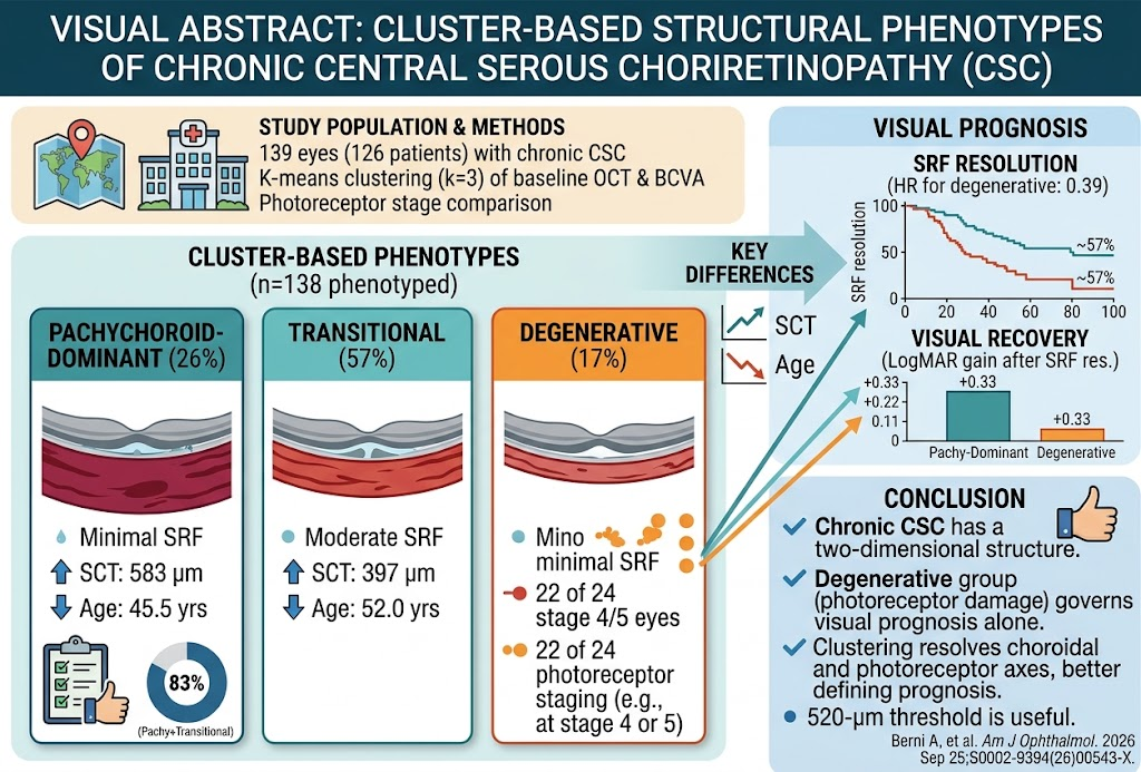
- Unsupervised k-means clustering of OCT-derived measurements and visual function identifies three distinct structural phenotypes of chronic CSC: pachychoroid-dominant, transitional, and degenerative.
- The photoreceptor damage threshold identified by clustering closely aligns with established photoreceptor staging and governs visual recovery after therapy.
- A second dimension characterized by choroidal thickness and age distinguishes morphologically similar groups, elucidating disease heterogeneity not captured by photoreceptor staging alone.
- Visual prognosis primarily depends on photoreceptor integrity whereas structural subgrouping refines understanding of pathophysiology and may influence therapeutic expectations.
Background
Chronic central serous chorioretinopathy (CSC) is a disorder marked by persistent or recurrent serous retinal detachment due to choroidal vascular hyperpermeability and retinal pigment epithelium (RPE) dysfunction. It predominately affects middle-aged adults and leads to visual impairment, especially in chronic cases. The pathophysiology is multifactorial, involving pachychoroid-driven choroidal thickening and secondary photoreceptor dysfunction. Despite advances in optical coherence tomography (OCT) imaging and photoreceptor staging systems, clinical heterogeneity remains, complicating prognosis and management. Verteporfin photodynamic therapy (PDT) is an established treatment promoting subretinal fluid (SRF) resolution. However, visual recovery varies considerably across patients.
Characterization of phenotypes combining structural (OCT) and functional metrics may clarify disease mechanisms, improve prognosis, and guide personalized interventions. The study by Berni et al. (2026) innovatively applies unsupervised clustering to delineate meaningful CSC subtypes beyond traditional staging.
Key Content
Study Design and Methodology
The reported multicenter retrospective cohort involved 139 eyes from 126 patients with chronic CSC treated with verteporfin PDT across referral centers. A robust unsupervised K-means clustering algorithm (k=3) applied four baseline variables: photoreceptor staging based on a validated six-stage OCT grading, central subfield retinal thickness, subfoveal choroidal thickness (SCT), and best-corrected visual acuity (BCVA). Resultant clusters were validated internally using out-of-bag accuracy (0.87) and externally against an independent chronic CSC cohort (55 eyes).
Phenotypic Classification and Characteristics
The clustering yielded three phenotypes:
- Pachychoroid-Dominant (26%): Characterized by markedly increased SCT (mean ~583 µm), younger mean age (~45.5 years), relatively preserved photoreceptors, and better baseline and post-treatment BCVA. This group exhibited higher rates of complete SRF resolution (83%).
- Transitional (57%): Intermediate phenotypic features with moderate SCT (~397 µm), older age (~52 years), and similar photoreceptor staging as pachychoroid-dominant eyes but reduced choroidal thickness and less favorable outcomes—SRF resolution at 76%.
- Degenerative (17%): Defined primarily by advanced photoreceptor damage (most eyes at stages 4 or 5), lowest SCT, oldest age (~65 years in validation cohort), worst baseline and visual recovery post-PDT, and the lowest SRF resolution rate (57%). This group exhibited a 0.33 logMAR less visual improvement than pachychoroid-dominant eyes after adjusting for baseline BCVA.
Notably, the phenotypic classification delineated a two-dimensional disease structure: one axis representing photoreceptor integrity directly linked to visual prognosis, and a second axis encompassing choroidal thickness and age, which photoreceptor staging alone failed to capture.
Comparative Analysis with Photoreceptor Staging
Although phenotypes largely paralleled photoreceptor staging for predicting visual outcomes, adding phenotype information did not significantly enhance predictive accuracy beyond photoreceptor stage alone for visual recovery or SRF resolution. Interestingly, pachychoroid-dominant and transitional eyes were indistinguishable by photoreceptor grade but separated by SCT and age, highlighting additional disease heterogeneity.
Validation in Independent Cohort
Phenotype proportions and the age gradient between groups were reproduced in an independent chronic CSC cohort, confirming external validity. The difference in choroidal thickness between groups was reduced but preserved the overall trend.
Clinical Outcomes and Implications
The degenerative group demonstrated significantly lower odds of achieving complete SRF resolution and poorer visual recovery post-therapy. The calibrated SCT threshold (~520 µm) was pivotal in distinguishing phenotypes, affirming the role of pachychoroid as a critical factor in disease classification. This stratification has potential to refine clinical prognosis and therapeutic counseling.
Expert Commentary
Chronic CSC remains a challenging clinical entity due to its variable course and outcome. The implementation of data-driven clustering methods leveraging multimodal OCT and functional data represents a methodological advance, enabling identification of biologically and clinically meaningful subtypes.
The study reconfirms the centrality of photoreceptor damage as the primary determinant of visual recovery, consistent with prior findings associating ellipsoid zone integrity and external limiting membrane status with functional prognosis. However, the novel identification of a choroidal thickness and age-related dimension expands our understanding of chronic CSC heterogeneity, echoing the pachychoroid concept’s evolving role.
Although phenotype assignment did not independently add prognostic value beyond photoreceptor classification for visual recovery, recognizing this second axis elucidates mechanistic nuances, such as differential choroidal remodeling and age-related changes influencing treatment responses and disease progression.
The relatively high cutoff SCT (~520 µm) calibrated to pachychoroid-confirmed eyes provides a practical biomarker for clinicians to subclassify patients and tailor management expectations. Given the suboptimal SRF resolution rates and visual gains in degenerative phenotypes, alternative or adjunctive therapeutic strategies may be warranted in this subgroup.
Limitations include retrospective design, potential selection bias from PDT-treated cohorts, and attenuation of choroidal thickness differences in validation cohorts, which may reflect variability in imaging or disease chronicity. Prospective studies incorporating functional imaging and biomarkers will be valuable to further refine CSC phenotyping and personalized treatment algorithms.
Conclusion
Chronic central serous chorioretinopathy exhibits a complex yet definable two-dimensional structural phenotype landscape. Unsupervised learning methods successfully recover the photoreceptor threshold critical for visual prognosis and reveal an orthogonal choroidal and age-related axis not captured by standard photoreceptor staging alone.
This refined phenotypic framework enhances disease stratification, potentially guiding clinical decision-making and improving individualized prognosis. Future research should validate these clusters longitudinally, explore underlying molecular pathways distinguishing phenotypes, and assess genotype-phenotype correlations to deepen mechanistic insights and optimize therapeutic approaches.
References
- Berni A, Scalabrin G, Neri G, et al. Cluster-Based Structural Phenotypes of Chronic Central Serous Chorioretinopathy. Am J Ophthalmol. 2026 Sep 25. PMID: 42790764.
- Daruich A, Matet A, Dirani A, et al. Central serous chorioretinopathy: recent findings and new physiopathology hypothesis. Prog Retin Eye Res. 2015;48:82-118. PMID: 26228067.
- Fujihara-Mino A, Yamashiro K, Ooto S, et al. Relationship between photoreceptor integrity and visual function in chronic central serous chorioretinopathy. Br J Ophthalmol. 2016;100(8):1149-1154. PMID: 26811507.
- Maruko I, Koizumi H, Nakashizuka H, et al. Subfoveal choroidal thickness in central serous chorioretinopathy. Retina. 2011;31(8):1445-1450. PMID: 21317874.
- Lim JW, Moon HJ, Choi H, et al. Photodynamic Therapy for Chronic Central Serous Chorioretinopathy: Meta-Analysis of Randomized Controlled Trials. Retina. 2021;41(7):1305-1314. PMID: 32957564.
This article was created using several editorial tools, including AI, as part of the process. Human editors reviewed this content before publication.