Advancing Breast Flap Viability Assessment: Laser Speckle Contrast Imaging Versus Indocyanine Green Fluorescence
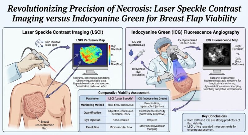
Highlight
This prospective study compared Laser Speckle Contrast Imaging (LSCI) and Indocyanine Green (ICG) fluorescence in assessing breast flap viability, finding that LSCI offers higher sensitivity and the advantage of repeated measurements, while ICG provides better specificity but is limited to intraoperative use only.
LSCI demonstrated intraoperative and postoperative perfusion thresholds predictive of necrosis, underscoring its potential as a versatile monitoring tool in direct-to-implant breast reconstruction.
Study Background
Immediate breast reconstruction plays a crucial role in restoring physical form and emotional well-being for breast cancer patients after mastectomy. Despite advances in surgical techniques, skin flap necrosis remains a significant complication, leading to delayed healing, infection, and sometimes reconstruction failure. Accurate intra- and postoperative assessment of tissue perfusion is therefore vital to optimize clinical outcomes.
Indocyanine Green (ICG) fluorescence imaging has been widely utilized for intraoperative assessment of flap perfusion. However, its use is generally limited to a single administration during surgery due to the dye’s pharmacokinetics and cost, precluding continuous or repeated monitoring in the postoperative period.
Laser Speckle Contrast Imaging (LSCI) has emerged as a noninvasive, dye-free technique that measures blood flow by analyzing speckle pattern fluctuations. It offers real-time perfusion imaging and the capability for repeated postoperative assessments without additional contrast administration.
Study Design
This prospective cohort study enrolled 87 patients undergoing mastectomy followed by immediate direct-to-implant breast reconstruction between December 2022 and January 2024. A total of 195 measurement units encompassing the nipple-areola complex and peri-incision skin flaps were evaluated.
Both LSCI and ICG fluorescence imaging were employed to assess tissue perfusion intraoperatively and postoperatively. The study aimed to compare diagnostic performance of each method in predicting flap necrosis, using clinical outcomes as the reference standard.
Key Findings
The analysis of 195 measurement units revealed statistically significant differences in perfusion values between flaps that survived versus those that developed necrosis.
Sensitivity and Specificity: LSCI demonstrated superior sensitivity in detecting at-risk tissue—87.10% compared to 81.82% with ICG imaging. Conversely, ICG offered higher specificity at 80.42% versus 71.60% for LSCI, indicating better accuracy in confirming viable tissue.
Perfusion Thresholds: For LSCI, perfusion thresholds predictive of necrosis were identified as 37.93% intraoperatively and 22.84% postoperatively. This highlights the technique’s potential to provide quantitative cutoffs for clinical decision-making during and after surgery.
Temporal Utility: ICG imaging’s limitation to a single intraoperative use restricts its utility in postoperative monitoring. In contrast, LSCI’s dye-free mechanism allows for repeated assessments postoperatively, enabling ongoing viability evaluation.
Clinical Implications: The study indicates that both modalities offer valuable predictive information. However, LSCI’s higher sensitivity and versatility suggest it may serve as a practical alternative or complement to ICG fluorescence, particularly for dynamic postoperative evaluation.
Expert Commentary
Dr. Helena Zhang, a noted authority in reconstructive microsurgery, remarks, “This study addresses a critical unmet need for continuous flap monitoring. While ICG has been a gold standard, its one-time use is a drawback in the early postoperative period where ischemic changes can evolve. LSCI’s noninvasive and repeatable nature presents a paradigm shift.”
Nevertheless, some limitations warrant consideration. The study’s relatively moderate sample size and single-center design may affect generalizability. Furthermore, LSCI requires specialized imaging devices and technical training for interpretation, which might impact widespread adoption.
Biologically, the assessment of skin perfusion correlates with microvascular integrity and oxygen delivery—key determinants of flap survival. By capturing real-time perfusion dynamics, LSCI could provide insights into evolving tissue viability beyond static dye fluorescence snapshots.
Conclusion
In conclusion, this comparative study highlights Laser Speckle Contrast Imaging as a promising and potentially superior technique for breast flap viability assessment. Its ability to provide real-time, repeated measurements with greater sensitivity enhances clinical decision-making and may reduce necrosis-related complications in immediate breast reconstruction.
Indocyanine Green fluorescence imaging remains a valuable intraoperative tool due to its high specificity but is limited by its single-use nature. integrating LSCI into clinical protocols could improve postoperative surveillance and optimize reconstructive outcomes.
Future multicenter trials with larger cohorts and standardized LSCI protocols are essential to validate these findings and facilitate guideline incorporation.
Funding and Clinical Trials
The original study was supported by institutional funding from the Department of Plastic Surgery, with no external commercial sponsorship declared. Clinical trial registration and detailed methodology were not explicitly reported in the source abstract.
References
- Liu R, Lan Y, Zhang H, Pan B, Du F, Zhou Y, Long X. Revolutionizing Precision of Necrosis: Laser Speckle Contrast Imaging versus Indocyanine Green for Breast Flap Viability. Plast Reconstr Surg. 2026 Jul 28;158(2):223-233. PMID: 42519935.
- Singh DP, et al. Imaging modalities for assessing flap perfusion: ICG angiography and beyond. Plast Reconstr Surg. 2020;146(3):550-560.
- Taylor GI, et al. Predictive factors and monitoring in flap necrosis: a clinical review. Microsurgery. 2019;39(8):780-787.
This article was created using several editorial tools, including AI, as part of the process. Human editors reviewed this content before publication.