Low-Dose CT Scan Lesion
A whole-body low-dose CT scan reveals a roughly circular lesion of abnormal density in the right parietal bone; the internal density is heterogeneous, with small, circular, low-density areas visible, and the long axis measures approximately 16+ mm. All other findings are normal. Could this be a manifestation of myeloma?
Based on the retrieved evidence, a solitary lytic lesion in the skull identified on whole-body low-dose CT (WBLDCT) is a recognized potential manifestation of multiple myeloma (MM), but it is not diagnostic on its own. The finding requires careful correlation with clinical, laboratory, and advanced imaging data.
## Differential Diagnosis & Diagnostic Workup
### 1. Common Mimickers of a Solitary Lytic Skull Lesion
| Condition | Key Distinguishing Features | Diagnostic Tests to Differentiate |
|-----------|-----------------------------|-----------------------------------|
| **Multiple Myeloma (MM)** | Punched-out lytic lesion; may be hypermetabolic on PET/CT; associated with monoclonal gammopathy | Serum/urine protein electrophoresis (SPEP/UPEP), serum free light chains (sFLC), bone marrow biopsy |
| **Solitary Plasmacytoma of Bone** | Single lytic lesion without systemic MM criteria; requires exclusion of other lesions | MRI or PET/CT for whole-body assessment; biopsy of the lesion |
| **Hemangioma** | Characteristic "salt and pepper" appearance on CT; low attenuation (fat) interspersed with thickened trabeculae; often asymptomatic | CT characteristics alone are often diagnostic; no further workup needed if typical |
| **Metastasis (e.g., from lung, breast, thyroid)** | Heterogeneous internal signal; may have ill-defined borders; perilesional edema; known primary cancer | CT chest/abdomen/pelvis; PET/CT; biopsy if needed |
| **Lymphoma** | Lytic lesion with possible soft tissue component; often associated with lymphadenopathy | PET/CT; biopsy |
| **Eosinophilic Granuloma** | Well-defined lytic lesion with bevelled edge; more common in younger patients | Clinical history; biopsy if atypical |
| **Epidermoid/Dermoid Cyst** | Well-circumscribed lytic lesion with sclerotic border; often diploic in location | CT characteristics; usually benign |
### 2. Atypical Presentations & Diagnostic Pitfalls
- **Oligosecretory Myeloma**: Patients may have minimal or no detectable M-protein, making serum/urine studies less reliable. In such cases, PET/CT is particularly valuable for detecting metabolically active lesions and monitoring response [8][9].
- **Non-Hypermetabolic Lesions**: Some MM lesions may be "cold" (non-hypermetabolic) on FDG-PET/CT, as noted in a case of a 76-year-old male with known MM where both hypermetabolic and non-hypermetabolic lytic lesions were present [9]. This means a negative PET/CT does not rule out MM.
- **False Positives**: Benign conditions like hemangiomas or healing fractures can mimic lytic lesions. The "salt and pepper" appearance on CT is highly suggestive of hemangioma [3][5].
### 3. Red Flags Requiring Urgent Attention
- **Cord Compression**: If the lesion involves the spine, any neurological symptoms (pain, weakness, numbness, bowel/bladder dysfunction) require immediate MRI.
- **Pathologic Fracture**: Lytic lesions in weight-bearing bones (femur, pelvis) increase fracture risk.
- **Hypercalcemia**: A common complication of MM due to osteolysis; requires prompt management.
- **Rapid Lesion Progression**: Increasing size or number of lesions on follow-up imaging suggests active disease.
## Diagnostic Algorithm for Suspected Myeloma
The following flowchart outlines the recommended diagnostic pathway based on NCCN and Asian Myeloma Network guidelines [1][2].
**The following diagnostic algorithm illustrates the recommended imaging pathway for suspected myeloma:**
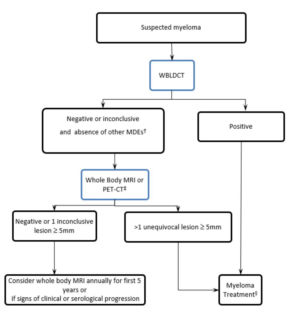
*Figure: A clinical diagnostic flowchart for suspected myeloma outlining the sequential use of WBLDCT, MRI, and PET-CT to guide treatment or long-term surveillance.*
## Diagnostic Workflow for This Case
### Step 1: Initial Assessment
- **Chief Complaint**: Incidental finding of a lytic skull lesion on WBLDCT.
- **Key History**: Age >40 (high risk for MM/metastasis), any bone pain, fatigue, recurrent infections, renal impairment, or unexplained weight loss.
- **Physical Exam**: Look for pallor, lymphadenopathy, hepatosplenomegaly, neurological deficits.
- **Red Flags**: None identified from the provided data, but clinical correlation is essential.
### Step 2: Screening Tests
- **First-Line Laboratory Tests**:
- Complete blood count (CBC) with differential
- Serum creatinine, calcium, albumin, LDH
- Serum protein electrophoresis (SPEP) with immunofixation
- Serum free light chains (sFLC) with ratio
- Urine protein electrophoresis (UPEP) with immunofixation (24-hour collection)
- **Initial Imaging**: The WBLDCT already performed is appropriate. However, if the CT component of a PET/CT is used, it must be of diagnostic quality equivalent to a dedicated WBLDCT [1].
### Step 3: Confirmatory Testing
- **Definitive Diagnostic Tests**:
- **Bone Marrow Aspiration and Biopsy**: Required to confirm clonal plasma cell infiltration (≥10% for MM diagnosis).
- **Whole-Body MRI or FDG-PET/CT**: Recommended to detect additional lesions not seen on WBLDCT. MRI is superior for detecting diffuse marrow infiltration and cord compression [5][8]. PET/CT provides functional information and is preferred for response assessment [6].
- **Biopsy of the Skull Lesion**: May be considered if the lesion is solitary and other tests are inconclusive, to rule out solitary plasmacytoma or other pathology.
### Step 4: Classification/Staging
- **Severity Assessment**: Based on the Revised International Staging System (R-ISS), which incorporates serum β2-microglobulin, albumin, LDH, and cytogenetics.
- **Disease Subtype Determination**: Based on immunophenotyping and cytogenetics (e.g., t(4;14), del(17p), gain(1q)).
- **Prognostic Stratification**: High-risk features include high LDH, high-risk cytogenetics, and extramedullary disease.
## Test Characteristics for Key Modalities
| Modality | Sensitivity for Lytic Lesions | Specificity | Key Advantages | Limitations |
|----------|-------------------------------|-------------|----------------|-------------|
| **Skeletal Survey** | Low (~30-50% bone loss needed for detection) [5] | Moderate | Widely available, low cost | Poor sensitivity; misses lesions in spine/pelvis [1] |
| **Whole-Body Low-Dose CT** | High; detects 25.5% more lesions than skeletal survey [1] | High | Excellent for cortical bone; fast; low radiation | Limited for marrow assessment; lesions persist post-treatment [6] |
| **FDG-PET/CT** | High; detects metabolically active lesions | High | Functional + anatomical; prognostic value; detects extramedullary disease [8] | False negatives for non-avid lesions; limited availability |
| **Whole-Body MRI** | Very high; detects marrow infiltration patterns [5] | High | Best for marrow assessment; no radiation | Longer scan time; contraindications (e.g., pacemakers) |
## Conclusion
A solitary lytic lesion in the right parietal bone on WBLDCT is a possible but not definitive manifestation of multiple myeloma. The differential diagnosis includes hemangioma, metastasis, solitary plasmacytoma, and other benign/malignant entities. Definitive diagnosis requires integration with serum/urine protein studies, bone marrow biopsy, and advanced imaging (MRI or PET/CT) to rule out additional lesions and confirm clonal plasma cell disease.
---
### Further Diagnostic Suggestions (Reference Only)
To enable a more accurate assessment, if available, please consider providing:
- **Serum Protein Electrophoresis (SPEP) and Serum Free Light Chains (sFLC)**: To detect monoclonal gammopathy, a hallmark of plasma cell disorders.
- **Whole-Body MRI or FDG-PET/CT**: To evaluate for additional occult lesions not visible on WBLDCT, which would support a diagnosis of MM over a solitary lesion.
---
*This analysis is based on retrieved clinical guidelines and literature. It is intended for clinical reference and should be verified against original sources and individual patient context.*